当前位置:
X-MOL 学术
›
J. Neurosci. Res.
›
论文详情
Our official English website, www.x-mol.net, welcomes your feedback! (Note: you will need to create a separate account there.)
The ependymal cell cytoskeleton in the normal and injured spinal cord of mice
Journal of Neuroscience Research ( IF 4.2 ) Pub Date : 2021-07-20 , DOI: 10.1002/jnr.24918 Omar Trujillo-Cenóz 1 , María I Rehermann 1 , Cecilia Maciel 1 , María V Falco 1 , Gabriela Fabbiani 1 , Raúl E Russo 1
Journal of Neuroscience Research ( IF 4.2 ) Pub Date : 2021-07-20 , DOI: 10.1002/jnr.24918 Omar Trujillo-Cenóz 1 , María I Rehermann 1 , Cecilia Maciel 1 , María V Falco 1 , Gabriela Fabbiani 1 , Raúl E Russo 1
Affiliation
|
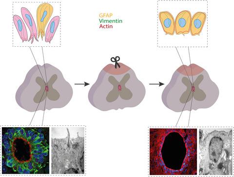
The cytoskeleton of ependymal cells is fundamental to organize and maintain the normal architecture of the central canal (CC). However, little is known about the plasticity of cytoskeletal components after spinal cord injury. Here, we focus on the structural organization of the cytoskeleton of ependymal cells in the normal and injured spinal cord of mice (both females and males) using immunohistochemical and electron microscopy techniques. We found that in uninjured animals, the actin cytoskeleton (as revealed by phalloidin staining) was arranged following the typical pattern of polarized epithelial cells with conspicuous actin pools located in the apical domain of ependymal cells. Transmission electron microscopy images showed microvilli tufts, long cilia, and characteristic intercellular membrane specializations. After spinal cord injury, F-actin rearrangements paralleled by fine structural modifications of the apical domain of ependymal cells were observed. These changes involved disruptions of the apical actin pools as well as fine structural modifications of the microvilli tufts. When comparing the control and injured spinal cords, we also found modifications in the expression of vimentin and glial fibrillary acidic protein (GFAP). After injury, vimentin expression disappeared from the most apical domains of ependymal cells but the number of GFAP-expressing cells within the CC increased. As in other polarized epithelia, the plastic changes in the cytoskeleton may be critically involved in the reaction of ependymal cells following a traumatic injury of the spinal cord.
中文翻译:

小鼠正常和损伤脊髓的室管膜细胞骨架
室管膜细胞的细胞骨架是组织和维持中央管 (CC) 正常结构的基础。然而,关于脊髓损伤后细胞骨架成分的可塑性知之甚少。在这里,我们使用免疫组织化学和电子显微镜技术关注小鼠(雌性和雄性)正常和受伤脊髓中室管膜细胞的细胞骨架的结构组织。我们发现在未受伤的动物中,肌动蛋白细胞骨架(如鬼笔环肽染色所示)按照极化上皮细胞的典型模式排列,在室管膜细胞的顶端区域具有明显的肌动蛋白池。透射电子显微镜图像显示微绒毛簇、长纤毛和特征性的细胞间膜特化。脊髓损伤后,观察到 F-肌动蛋白重排与室管膜细胞顶端结构域的精细结构修饰平行。这些变化涉及顶端肌动蛋白池的破坏以及微绒毛簇的精细结构修饰。在比较对照组和受伤脊髓时,我们还发现波形蛋白和胶质纤维酸性蛋白 (GFAP) 的表达发生了变化。损伤后,波形蛋白表达从室管膜细胞的最顶端区域消失,但 CC 内表达 GFAP 的细胞数量增加。与其他极化上皮细胞一样,细胞骨架的可塑性变化可能与脊髓外伤后室管膜细胞的反应密切相关。
更新日期:2021-07-20
中文翻译:
小鼠正常和损伤脊髓的室管膜细胞骨架
室管膜细胞的细胞骨架是组织和维持中央管 (CC) 正常结构的基础。然而,关于脊髓损伤后细胞骨架成分的可塑性知之甚少。在这里,我们使用免疫组织化学和电子显微镜技术关注小鼠(雌性和雄性)正常和受伤脊髓中室管膜细胞的细胞骨架的结构组织。我们发现在未受伤的动物中,肌动蛋白细胞骨架(如鬼笔环肽染色所示)按照极化上皮细胞的典型模式排列,在室管膜细胞的顶端区域具有明显的肌动蛋白池。透射电子显微镜图像显示微绒毛簇、长纤毛和特征性的细胞间膜特化。脊髓损伤后,观察到 F-肌动蛋白重排与室管膜细胞顶端结构域的精细结构修饰平行。这些变化涉及顶端肌动蛋白池的破坏以及微绒毛簇的精细结构修饰。在比较对照组和受伤脊髓时,我们还发现波形蛋白和胶质纤维酸性蛋白 (GFAP) 的表达发生了变化。损伤后,波形蛋白表达从室管膜细胞的最顶端区域消失,但 CC 内表达 GFAP 的细胞数量增加。与其他极化上皮细胞一样,细胞骨架的可塑性变化可能与脊髓外伤后室管膜细胞的反应密切相关。


 京公网安备 11010802027423号