当前位置:
X-MOL 学术
›
Brain Pathol.
›
论文详情
Our official English website, www.x-mol.net, welcomes your
feedback! (Note: you will need to create a separate account there.)
41-year-old male with a pituitary mass
Brain Pathology ( IF 5.8 ) Pub Date : 2021-07-19 , DOI: 10.1111/bpa.12961 Wenjuan Wen 1 , Leiming Wang 2 , Mengyou Li 3 , Peijin Li 1 , Yubo Ren 1 , Xuedong Zhang 1
Brain Pathology ( IF 5.8 ) Pub Date : 2021-07-19 , DOI: 10.1111/bpa.12961 Wenjuan Wen 1 , Leiming Wang 2 , Mengyou Li 3 , Peijin Li 1 , Yubo Ren 1 , Xuedong Zhang 1
Affiliation
|
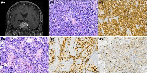
Cranial coronal T1-weighted magnetic resonance imaging with contrast enhancement showed a sellar irregular lesion (Figure A). Hematoxylin and eosin staining showed two different morphologies. The majority of tumor cells had medium-sized to large cells with a high nucleus to cytoplasm ratio, vesicular nuclei with prominent nucleoli, and poor adhesion (Figure B), which revealed positive expression of CD20 by Immunohistochemistry (Figure C). The other component showed abundant cytoplasm, spindle-like to ovoid nucleus and rare mitotic figures (Figure D). These tumor cells were positive for Pit-1 (Figure E) and perinuclear punctated structures immunopositive for CK18 (Figure F).

中文翻译:

41岁男性垂体肿块
头颅冠状 T1 加权磁共振成像对比增强显示鞍区不规则病变(图 A)。苏木精和伊红染色显示两种不同的形态。大部分肿瘤细胞为中至大细胞,核质比高,胞核呈水泡状,核仁明显,粘附性差(图B),免疫组化显示CD20阳性表达(图C)。其他成分显示丰富的细胞质,梭形至卵圆形核和罕见的有丝分裂图(图D)。这些肿瘤细胞对 Pit-1 呈阳性(图 E)和对 CK18 呈免疫阳性的核周点状结构(图 F)。
更新日期:2021-07-19
中文翻译:
41岁男性垂体肿块
头颅冠状 T1 加权磁共振成像对比增强显示鞍区不规则病变(图 A)。苏木精和伊红染色显示两种不同的形态。大部分肿瘤细胞为中至大细胞,核质比高,胞核呈水泡状,核仁明显,粘附性差(图B),免疫组化显示CD20阳性表达(图C)。其他成分显示丰富的细胞质,梭形至卵圆形核和罕见的有丝分裂图(图D)。这些肿瘤细胞对 Pit-1 呈阳性(图 E)和对 CK18 呈免疫阳性的核周点状结构(图 F)。


 京公网安备 11010802027423号