Cellular and Molecular Neurobiology ( IF 3.6 ) Pub Date : 2021-06-09 , DOI: 10.1007/s10571-021-01116-0 Anna Hilverling 1 , Eva M Szegö 2 , Elisabeth Dinter 2 , Diana Cozma 2 , Theodora Saridaki 1 , Björn H Falkenburger 1, 2, 3, 4
|
Autophagosome maturation comprises fusion with lysosomes and acidification. It is a critical step in the degradation of cytosolic protein aggregates that characterize many neurodegenerative diseases. In order to better understand this process, we studied intracellular trafficking of autophagosomes and aggregates of α-synuclein, which characterize Parkinson’s disease and other synucleinopathies. The autophagosomal marker LC3 and the aggregation prone A53T mutant of α-synuclein were tagged by fluorescent proteins and expressed in HEK293T cells and primary astrocytes. The subcellular distribution and movement of these vesicle populations were analyzed by (time-lapse) microscopy. Fusion with lysosomes was assayed using the lysosomal marker LAMP1; vesicles with neutral and acidic luminal pH were discriminated using the RFP-GFP “tandem-fluorescence” tag. With respect to vesicle pH, we observed that neutral autophagosomes, marked by LC3 or synuclein, were located more frequently in the cell center, and acidic autophagosomes were observed more frequently in the cell periphery. Acidic autophagosomes were transported towards the cell periphery more often, indicating that acidification occurs in the cell center before transport to the periphery. With respect to autolysosomal fusion, we found that lysosomes preferentially moved towards the cell center, whereas autolysosomes moved towards the cell periphery, suggesting a cycle where lysosomes are generated in the periphery and fuse to autophagosomes in the cell center. Unexpectedly, many acidic autophagosomes were negative for LAMP1, indicating that acidification does not require fusion to lysosomes. Moreover, we found both neutral and acidic vesicles positive for LAMP1, consistent with delayed acidification of the autolysosome lumen. Individual steps of aggregate clearance thus occur in dedicated cellular regions. During aggregate clearance, autophagosomes and autolysosomes form in the center and are transported towards the periphery during maturation. In this process, luminal pH could regulate the direction of vesicle transport.
Graphic Abstract
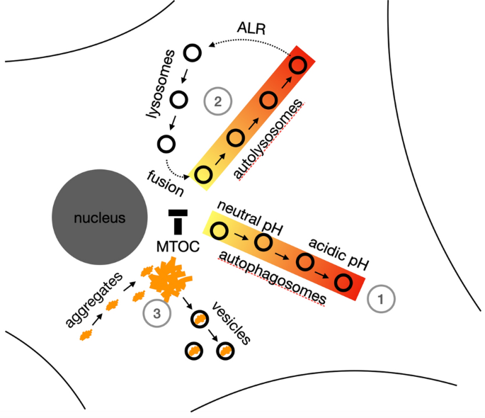
(1) Transport and location of autophagosomes depend on luminal pH: Acidic autophagosomes are preferentially transported to the cell periphery, causing more acidic autophagosomes in the cell periphery and more neutral autophagosomes at the microtubule organizing center (MTOC). (2) Autolysosomes are transported to the cell periphery and lysosomes to the MTOC, suggesting spatial segregation of lysosome reformation and autolysosome fusion. (3) Synuclein aggregates are preferentially located at the MTOC and synuclein-containing vesicles in the cell periphery, consistent with transport of aggregates to the MTOC for autophagy.
中文翻译:
成熟的自噬体被运送到细胞外围
自噬体成熟包括与溶酶体的融合和酸化。这是细胞溶质蛋白聚集体降解的关键步骤,这是许多神经退行性疾病的特征。为了更好地理解这一过程,我们研究了自噬体和 α-突触核蛋白聚集体的细胞内运输,这是帕金森病和其他突触核蛋白病的特征。自噬体标志物 LC3 和易聚集的 α-突触核蛋白 A53T 突变体被荧光蛋白标记并在 HEK293T 细胞和原代星形胶质细胞中表达。通过(延时)显微镜分析这些囊泡群的亚细胞分布和运动。使用溶酶体标记 LAMP1 检测与溶酶体的融合;使用 RFP-GFP“串联荧光”标签区分具有中性和酸性管腔 pH 值的囊泡。关于囊泡的 pH 值,我们观察到以 LC3 或突触核蛋白为标志的中性自噬体更频繁地位于细胞中心,而酸性自噬体更频繁地位于细胞外围。酸性自噬体更频繁地被转运到细胞外围,表明酸化发生在细胞中心,然后再转运到外围。关于自溶酶体融合,我们发现溶酶体优先向细胞中心移动,而自溶酶体向细胞外围移动,表明溶酶体在外围产生并与细胞中心的自噬体融合的循环。出乎意料的是,许多酸性自噬体对 LAMP1 呈阴性,表明酸化不需要与溶酶体融合。而且,我们发现中性和酸性囊泡都对 LAMP1 呈阳性,这与自溶酶体腔的延迟酸化一致。因此,聚集清除的各个步骤发生在专用的细胞区域。在聚集体清除过程中,自噬体和自溶酶体在中心形成,并在成熟过程中被转运到外围。在这个过程中,腔内的 pH 值可以调节囊泡运输的方向。
图形摘要
(1)自噬体的转运和定位取决于腔内pH值:酸性自噬体优先转运到细胞外周,导致细胞外周酸性自噬体增多,微管组织中心(MTOC)中性自噬体增多。(2)自溶酶体被转运到细胞外围,溶酶体被转运到MTOC,表明溶酶体重组和自溶酶体融合的空间分离。(3) 突触核蛋白聚集体优先位于细胞外周的 MTOC 和含有突触核蛋白的囊泡,这与将聚集体运输到 MTOC 进行自噬相一致。


 京公网安备 11010802027423号