当前位置:
X-MOL 学术
›
J. Neuroendocrinol.
›
论文详情
Our official English website, www.x-mol.net, welcomes your
feedback! (Note: you will need to create a separate account there.)
Increased neuronal activation in sympathoregulatory regions of the brain and spinal cord in type 2 diabetic rats
Journal of Neuroendocrinology ( IF 3.3 ) Pub Date : 2021-07-15 , DOI: 10.1111/jne.13016 Shivani Sethi 1, 2, 3, 4 , Rachael A Augustine 1, 2, 3, 4 , Gregory T Bouwer 1, 3, 4 , Michael R Perkinson 1, 3, 4 , Isaiah Cheong 1, 2, 3, 4 , Carol T Bussey 1, 2, 5 , Daryl O Schwenke 1, 2 , Colin H Brown 1, 3, 4 , Regis R Lamberts 1, 2
Journal of Neuroendocrinology ( IF 3.3 ) Pub Date : 2021-07-15 , DOI: 10.1111/jne.13016 Shivani Sethi 1, 2, 3, 4 , Rachael A Augustine 1, 2, 3, 4 , Gregory T Bouwer 1, 3, 4 , Michael R Perkinson 1, 3, 4 , Isaiah Cheong 1, 2, 3, 4 , Carol T Bussey 1, 2, 5 , Daryl O Schwenke 1, 2 , Colin H Brown 1, 3, 4 , Regis R Lamberts 1, 2
Affiliation
|
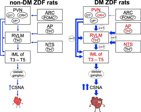
Increased cardiac sympathetic nerve activity in type 2 diabetes mellitus (DM) suggests impaired autonomic control of the heart. However, the central regions that contribute to the autonomic cardiac pathologies in type 2 DM are unknown. Therefore, we tested the hypothesis that neuronal activation would be increased in central sympathoregulatory areas in a pre-clinical type 2 DM animal model. Immunohistochemistry in 20-week-old male Zucker diabetic fatty (ZDF) rats revealed an increased number of neurones expressing ΔFosB (a marker of chronic neuronal activation) in the intermediolateral column (IML) of the spinal cord in DM compared to non-diabetic (non-DM) rats (P < 0.05). Rostral ventrolateral medulla (RVLM) neurones activate IML neurones and receive inputs from the hypothalamic paraventricular nucleus (PVN), as well as the nucleus tractus solitarius (NTS) and area postrema (AP), in the brainstem. We observed more ΔFosB-positive noradrenergic RVLM neurones (P < 0.001) and corticotrophin-releasing hormone PVN neurones (P < 0.05) in DM compared to non-DM rats. More ΔFosB-positive neurones were also observed in the NTS (P < 0.05) and AP (P < 0.01) of DM rats compared to non-DM rats. Finally, because DM ZDF rats are obese, we also expected increased activation of pro-opiomelanocortin (POMC) arcuate nucleus (ARC) neurones in DM rats; however, fewer ΔFosB-positive POMC ARC neurones were observed in DM compared to non-DM rats (P < 0.01). In conclusion, increased neuronal activation in the IML of type 2 DM ZDF rats might be driven by RVLM neurones that are possibly activated by PVN, NTS and AP inputs. Elucidating the contribution of central sympathoexcitatory drive in type 2 DM might improve the effectiveness of pharmacotherapies for diabetic heart disease.
中文翻译:

2 型糖尿病大鼠大脑和脊髓交感调节区神经元激活增加
2 型糖尿病 (DM) 患者心脏交感神经活动增加表明心脏自主控制受损。然而,导致 2 型糖尿病自主神经心脏病的中心区域尚不清楚。因此,我们在临床前 2 型糖尿病动物模型中检验了中枢交感神经调节区神经元激活会增加的假设。 20 周龄雄性 Zucker 糖尿病肥胖 (ZDF) 大鼠的免疫组织化学显示,与非糖尿病大鼠相比,DM 脊髓中外侧柱 (IML) 表达 ΔFosB(慢性神经元激活标志物)的神经元数量增加。非糖尿病)大鼠( P < 0.05)。延髓头侧腹外侧 (RVLM) 神经元激活 IML 神经元并接收来自下丘脑室旁核 (PVN) 以及脑干中孤束核 (NTS) 和后区 (AP) 的输入。与非糖尿病大鼠相比,我们在糖尿病大鼠中观察到更多的 ΔFosB 阳性去甲肾上腺素能 RVLM 神经元 ( P < 0.001) 和促肾上腺皮质激素释放激素 PVN 神经元 ( P < 0.05)。与非DM大鼠相比,DM大鼠的NTS( P <0.05)和AP( P <0.01)中也观察到更多的ΔFosB阳性神经元。最后,由于 DM ZDF 大鼠肥胖,我们还预计 DM 大鼠中阿片黑皮素原 (POMC) 弓状核 (ARC) 神经元的激活增加;然而,与非 DM 大鼠相比,DM 大鼠中观察到的 ΔFosB 阳性 POMC ARC 神经元较少( P < 0.01)。总之,2 型 DM ZDF 大鼠 IML 中神经元激活的增加可能是由 RVLM 神经元驱动的,而 RVLM 神经元可能由 PVN、NTS 和 AP 输入激活。 阐明中枢交感神经兴奋驱动在 2 型糖尿病中的作用可能会提高药物治疗糖尿病性心脏病的有效性。
更新日期:2021-09-10
中文翻译:
2 型糖尿病大鼠大脑和脊髓交感调节区神经元激活增加
2 型糖尿病 (DM) 患者心脏交感神经活动增加表明心脏自主控制受损。然而,导致 2 型糖尿病自主神经心脏病的中心区域尚不清楚。因此,我们在临床前 2 型糖尿病动物模型中检验了中枢交感神经调节区神经元激活会增加的假设。 20 周龄雄性 Zucker 糖尿病肥胖 (ZDF) 大鼠的免疫组织化学显示,与非糖尿病大鼠相比,DM 脊髓中外侧柱 (IML) 表达 ΔFosB(慢性神经元激活标志物)的神经元数量增加。非糖尿病)大鼠( P < 0.05)。延髓头侧腹外侧 (RVLM) 神经元激活 IML 神经元并接收来自下丘脑室旁核 (PVN) 以及脑干中孤束核 (NTS) 和后区 (AP) 的输入。与非糖尿病大鼠相比,我们在糖尿病大鼠中观察到更多的 ΔFosB 阳性去甲肾上腺素能 RVLM 神经元 ( P < 0.001) 和促肾上腺皮质激素释放激素 PVN 神经元 ( P < 0.05)。与非DM大鼠相比,DM大鼠的NTS( P <0.05)和AP( P <0.01)中也观察到更多的ΔFosB阳性神经元。最后,由于 DM ZDF 大鼠肥胖,我们还预计 DM 大鼠中阿片黑皮素原 (POMC) 弓状核 (ARC) 神经元的激活增加;然而,与非 DM 大鼠相比,DM 大鼠中观察到的 ΔFosB 阳性 POMC ARC 神经元较少( P < 0.01)。总之,2 型 DM ZDF 大鼠 IML 中神经元激活的增加可能是由 RVLM 神经元驱动的,而 RVLM 神经元可能由 PVN、NTS 和 AP 输入激活。 阐明中枢交感神经兴奋驱动在 2 型糖尿病中的作用可能会提高药物治疗糖尿病性心脏病的有效性。


 京公网安备 11010802027423号