Computer Methods and Programs in Biomedicine ( IF 4.9 ) Pub Date : 2021-07-14 , DOI: 10.1016/j.cmpb.2021.106265 Shiyu Wang 1 , Xiang Liu 1 , Jingwen Zhao 1 , Yiwen Liu 1 , Shuhong Liu 2 , Yisi Liu 2 , Jingmin Zhao 2
|
Background and objectives
Cholangiocarcinoma (CCA) is one of the most aggressive human malignant tumors and is becoming one of the main factors of death and disability globally. Specifically, 60% to 70% of CCA patients were diagnosed with local invasion or distant metastasis and lost the chance of radical operation. The overall median survival time was less than 12 months. As a non-invasive diagnostic technology, medical imaging consisting of computed tomography (CT) imaging, magnetic resonance imaging (MRI), and ultrasound (US) imaging, is the most effectively and commonly used method to detect CCA. The computer auxiliary diagnosis (CAD) system based on medical imaging is helpful for rapid diagnosis and provides credible “second opinion” for specialists. The purpose of this review is to categorize and review the CAD technique of detecting CCA based on medical imaging.
Methods
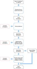
This work applies a four-level screening process to choose suitable publications. 125 research papers published in different academic research databases were selected and analyzed according to specific criteria. From the five steps of medical image acquisition, processing, analysis, understanding and verification of CAD combined with artificial intelligence algorithms, we obtain the most advanced insights related to CCA detection.
Results
This work provides a comprehensive analysis and comparison analysis of the current CAD systems of detecting CCA. After careful investigation, we find that the main detection methods are traditional machine learning method and deep learning method. For the detection, the most commonly used method is semi-automatic segmentation algorithm combined with support vector machine classifier method, combination of which has good detection performance. The end-to-end training mode makes deep learning method more and more popular in CAD systems. However, due to the limited medical training data, the accuracy of deep learning method is unsatisfactory.
Conclusions
Based on analysis of artificial intelligence methods applied in CCA, this work is expected to be truly applied in clinical practice in the future to improve the level of clinical diagnosis and treatment of it. This work concludes by providing a prediction of future trends, which will be of great significance for researchers in the medical imaging of CCA and artificial intelligence.
中文翻译:
基于医学影像检测胆管癌的计算机辅助诊断技术综述
背景和目标
胆管癌(CCA)是最具侵袭性的人类恶性肿瘤之一,正在成为全球死亡和残疾的主要因素之一。具体而言,60%~70%的CCA患者被诊断为局部浸润或远处转移,失去了根治性手术的机会。总体中位生存时间小于 12 个月。作为一种无创诊断技术,由计算机断层扫描(CT)成像、磁共振成像(MRI)和超声(US)成像组成的医学成像是检测CCA最有效和最常用的方法。基于医学影像的计算机辅助诊断(CAD)系统有助于快速诊断,为专家提供可信的“第二意见”。本综述的目的是对基于医学影像检测 CCA 的 CAD 技术进行分类和回顾。
方法
这项工作采用四级筛选过程来选择合适的出版物。125篇发表在不同学术研究数据库中的研究论文被按照特定标准筛选和分析。从医学图像采集、处理、分析、理解和验证CAD的五个步骤结合人工智能算法,我们获得了与CCA检测相关的最先进的见解。
结果
这项工作对当前检测 CCA 的 CAD 系统进行了全面的分析和比较分析。经过仔细调查,我们发现主要的检测方法是传统的机器学习方法和深度学习方法。对于检测,最常用的方法是半自动分割算法结合支持向量机分类器方法,两者结合具有良好的检测性能。端到端的训练模式使得深度学习方法在 CAD 系统中越来越流行。然而,由于医学训练数据有限,深度学习方法的准确性并不尽如人意。
结论
基于人工智能方法在CCA中的应用分析,该工作有望在未来真正应用于临床,提高临床诊疗水平。这项工作最后提供了对未来趋势的预测,这对 CCA 和人工智能医学成像的研究人员具有重要意义。


 京公网安备 11010802027423号