当前位置:
X-MOL 学术
›
NMR Biomed.
›
论文详情
Our official English website, www.x-mol.net, welcomes your
feedback! (Note: you will need to create a separate account there.)
Utility of diffusion MRI characteristics of cervical lymph nodes as disease classifier between patients with head and neck squamous cell carcinoma and healthy volunteers
NMR in Biomedicine ( IF 2.7 ) Pub Date : 2021-07-08 , DOI: 10.1002/nbm.4587 Marianthi-Vasiliki Papoutsaki 1 , Harbir Singh Sidhu 1 , Nikolaos Dikaios 2 , Saurabh Singh 1 , David Atkinson 1 , Baris Kanber 3 , Timothy Beale 4 , Simon Morley 4 , Martin Forster 5, 6 , Dawn Carnell 6 , Ruheena Mendes 6 , Shonit Punwani 1
NMR in Biomedicine ( IF 2.7 ) Pub Date : 2021-07-08 , DOI: 10.1002/nbm.4587 Marianthi-Vasiliki Papoutsaki 1 , Harbir Singh Sidhu 1 , Nikolaos Dikaios 2 , Saurabh Singh 1 , David Atkinson 1 , Baris Kanber 3 , Timothy Beale 4 , Simon Morley 4 , Martin Forster 5, 6 , Dawn Carnell 6 , Ruheena Mendes 6 , Shonit Punwani 1
Affiliation
|
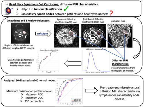
Diffusion MRI characteristics assessed by apparent diffusion coefficient (ADC) histogram analysis in head and neck squamous cell carcinoma (HNSCC) have been reported as helpful in classifying tumours based on diffusion characteristics. There is little reported on HNSCC lymph nodes classification by diffusion characteristics. The aim of this study was to determine whether pretreatment nodal microstructural diffusion MRI characteristics can classify diseased nodes of patients with HNSCC from normal nodes of healthy volunteers. Seventy-nine patients with histologically confirmed HNSCC prior to chemoradiotherapy, and eight healthy volunteers, underwent diffusion-weighted (DW) MRI at a 1.5-T MR scanner. Two radiologists contoured lymph nodes on DW (b = 300 s/m2) images. ADC, distributed diffusion coefficient (DDC) and alpha (α) values were calculated by monoexponential and stretched exponential models. Histogram analysis metrics of drawn volume were compared between patients and volunteers using a Mann–Whitney test. The classification performance of each metric between the normal and diseased nodes was determined by receiver operating characteristic (ROC) analysis. Intraclass correlation coefficients determined interobserver reproducibility of each metric based on differently drawn ROIs by two radiologists. Sixty cancerous and 40 normal nodes were analysed. ADC histogram analysis revealed significant differences between patients and volunteers (p ≤0.0001 to 0.0046), presenting ADC distributions that were more skewed (1.49 for patients, 1.03 for volunteers; p = 0.0114) and ‘peaked’ (6.82 for patients, 4.20 for volunteers; p = 0.0021) in patients. Maximum ADC values exhibited the highest area under the curve ([AUC] 0.892). Significant differences were revealed between patients and volunteers for DDC and α value histogram metrics (p ≤0.0001 to 0.0044); the highest AUC were exhibited by maximum DDC (0.772) and the 25th percentile α value (0.761). Interobserver repeatability was excellent for mean ADC (ICC = 0.88) and the 25th percentile α value (ICC = 0.78), but poor for all other metrics. These results suggest that pretreatment microstructural diffusion MRI characteristics in lymph nodes, assessed by ADC and α value histogram analysis, can identify nodal disease.
中文翻译:

颈部淋巴结弥散 MRI 特征作为头颈部鳞状细胞癌患者与健康志愿者疾病分类的效用
据报道,通过头颈部鳞状细胞癌 (HNSCC) 的表观扩散系数 (ADC) 直方图分析评估的扩散 MRI 特征有助于根据扩散特征对肿瘤进行分类。根据扩散特征对 HNSCC 淋巴结分类的报道很少。本研究的目的是确定治疗前淋巴结微结构弥散 MRI 特征是否可以将 HNSCC 患者的病变淋巴结与健康志愿者的正常淋巴结区分开来。放化疗前组织学证实为 HNSCC 的 79 名患者和 8 名健康志愿者在 1.5-T MR 扫描仪上接受了弥散加权 (DW) MRI。两位放射科医生在 DW 上勾勒出淋巴结轮廓 (b = 300 s/m 2) 图片。ADC、分布扩散系数 (DDC) 和 α (α) 值通过单指数和拉伸指数模型计算。使用 Mann-Whitney 检验比较患者和志愿者之间抽取体积的直方图分析指标。正常和患病节点之间的每个度量的分类性能由接收器操作特征 (ROC) 分析确定。组内相关系数根据两位放射科医生绘制的不同 ROI 确定了每个指标的观察者间可重复性。分析了 60 个癌性淋巴结和 40 个正常淋巴结。ADC 直方图分析显示患者和志愿者之间存在显着差异(p ≤0.0001 至 0.0046),表明 ADC 分布更加偏斜(患者为 1.49,志愿者为 1.03;p = 0.0114)和“峰值”(患者为 6.82,志愿者为 4.20;p = 0.0021)。最大 ADC 值表现出曲线下的最高面积 ([AUC] 0.892)。DDC 和 α 值直方图指标在患者和志愿者之间存在显着差异(p ≤0.0001 至 0.0044);最大 DDC (0.772) 和第 25 个百分位 α 值 (0.761) 表现出最高的 AUC。平均 ADC (ICC = 0.88) 和第 25 个百分位 α 值 (ICC = 0.78) 的观察者间重复性非常好,但对于所有其他指标来说都很差。这些结果表明,通过 ADC 和 α 值直方图分析评估的淋巴结的预处理显微结构弥散 MRI 特征可以识别淋巴结疾病。
更新日期:2021-07-08
中文翻译:
颈部淋巴结弥散 MRI 特征作为头颈部鳞状细胞癌患者与健康志愿者疾病分类的效用
据报道,通过头颈部鳞状细胞癌 (HNSCC) 的表观扩散系数 (ADC) 直方图分析评估的扩散 MRI 特征有助于根据扩散特征对肿瘤进行分类。根据扩散特征对 HNSCC 淋巴结分类的报道很少。本研究的目的是确定治疗前淋巴结微结构弥散 MRI 特征是否可以将 HNSCC 患者的病变淋巴结与健康志愿者的正常淋巴结区分开来。放化疗前组织学证实为 HNSCC 的 79 名患者和 8 名健康志愿者在 1.5-T MR 扫描仪上接受了弥散加权 (DW) MRI。两位放射科医生在 DW 上勾勒出淋巴结轮廓 (b = 300 s/m 2) 图片。ADC、分布扩散系数 (DDC) 和 α (α) 值通过单指数和拉伸指数模型计算。使用 Mann-Whitney 检验比较患者和志愿者之间抽取体积的直方图分析指标。正常和患病节点之间的每个度量的分类性能由接收器操作特征 (ROC) 分析确定。组内相关系数根据两位放射科医生绘制的不同 ROI 确定了每个指标的观察者间可重复性。分析了 60 个癌性淋巴结和 40 个正常淋巴结。ADC 直方图分析显示患者和志愿者之间存在显着差异(p ≤0.0001 至 0.0046),表明 ADC 分布更加偏斜(患者为 1.49,志愿者为 1.03;p = 0.0114)和“峰值”(患者为 6.82,志愿者为 4.20;p = 0.0021)。最大 ADC 值表现出曲线下的最高面积 ([AUC] 0.892)。DDC 和 α 值直方图指标在患者和志愿者之间存在显着差异(p ≤0.0001 至 0.0044);最大 DDC (0.772) 和第 25 个百分位 α 值 (0.761) 表现出最高的 AUC。平均 ADC (ICC = 0.88) 和第 25 个百分位 α 值 (ICC = 0.78) 的观察者间重复性非常好,但对于所有其他指标来说都很差。这些结果表明,通过 ADC 和 α 值直方图分析评估的淋巴结的预处理显微结构弥散 MRI 特征可以识别淋巴结疾病。


 京公网安备 11010802027423号