当前位置:
X-MOL 学术
›
Microsc. Res. Tech.
›
论文详情
Our official English website, www.x-mol.net, welcomes your
feedback! (Note: you will need to create a separate account there.)
Automatic detection of papilledema through fundus retinal images using deep learning
Microscopy Research and Technique ( IF 2.0 ) Pub Date : 2021-07-08 , DOI: 10.1002/jemt.23865 Tanzila Saba 1 , Shahzad Akbar 2 , Hoshang Kolivand 3, 4 , Saeed Ali Bahaj 5
Microscopy Research and Technique ( IF 2.0 ) Pub Date : 2021-07-08 , DOI: 10.1002/jemt.23865 Tanzila Saba 1 , Shahzad Akbar 2 , Hoshang Kolivand 3, 4 , Saeed Ali Bahaj 5
Affiliation
|
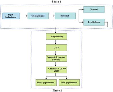
Papilledema is a syndrome of the retina in which retinal optic nerve is inflated by elevation of intracranial pressure. The papilledema abnormalities such as retinal nerve fiber layer (RNFL) opacification may lead to blindness. These abnormalities could be seen through capturing of retinal images by means of fundus camera. This paper presents a deep learning-based automated system that detects and grades the papilledema through U-Net and Dense-Net architectures. The proposed approach has two main stages. First, optic disc and its surrounding area in fundus retinal image are localized and cropped for input to Dense-Net which classifies the optic disc as papilledema or normal. Second, consists of preprocessing of Dense-Net classified papilledema fundus image by Gabor filter. The preprocessed papilledema image is input to U-Net to achieve the segmented vascular network from which the vessel discontinuity index (VDI) and vessel discontinuity index to disc proximity (VDIP) are calculated for grading of papilledema. The VDI and VDIP are standard parameter to check the severity and grading of papilledema. The proposed system is evaluated on 60 papilledema and 40 normal fundus images taken from STARE dataset. The experimental results for classification of papilledema through Dense-Net are much better in terms of sensitivity 98.63%, specificity 97.83%, and accuracy 99.17%. Similarly, the grading results for mild and severe papilledema classification through U-Net are also much better in terms of sensitivity 99.82%, specificity 98.65%, and accuracy 99.89%. The deep learning-based automated detection and grading of papilledema for clinical purposes is first effort in state of art.
中文翻译:

使用深度学习通过眼底视网膜图像自动检测视乳头水肿
视乳头水肿是一种视网膜综合征,其中视网膜视神经因颅内压升高而膨胀。视乳头水肿异常,如视网膜神经纤维层 (RNFL) 混浊,可能导致失明。这些异常可以通过眼底相机捕获视网膜图像来观察。本文提出了一种基于深度学习的自动化系统,该系统通过 U-Net 和 Dense-Net 架构检测和分级视乳头水肿。建议的方法有两个主要阶段。首先,眼底视网膜图像中的视盘及其周围区域被定位并裁剪以输入到 Dense-Net,该 Dense-Net 将视盘分类为视乳头水肿或正常。其次,由 Gabor 滤波器对 Dense-Net 分类的眼底视乳头图像进行预处理。将预处理后的视乳头水肿图像输入到 U-Net 以实现分割的血管网络,从中计算血管不连续指数 (VDI) 和血管不连续指数与椎间盘接近度 (VDIP),用于对视乳头水肿进行分级。VDI 和 VDIP 是检查视乳头水肿严重程度和分级的标准参数。所提出的系统在 60 个视乳头水肿和 40 个来自 STARE 数据集的正常眼底图像上进行评估。通过 Dense-Net 对视乳头水肿进行分类的实验结果在灵敏度 98.63%、特异性 97.83% 和准确度 99.17% 方面要好得多。同样,通过 U-Net 对轻度和重度视乳头水肿分类的分级结果在敏感性 99.82%、特异性 98.65% 和准确度 99.89% 方面也更好。
更新日期:2021-07-08
中文翻译:
使用深度学习通过眼底视网膜图像自动检测视乳头水肿
视乳头水肿是一种视网膜综合征,其中视网膜视神经因颅内压升高而膨胀。视乳头水肿异常,如视网膜神经纤维层 (RNFL) 混浊,可能导致失明。这些异常可以通过眼底相机捕获视网膜图像来观察。本文提出了一种基于深度学习的自动化系统,该系统通过 U-Net 和 Dense-Net 架构检测和分级视乳头水肿。建议的方法有两个主要阶段。首先,眼底视网膜图像中的视盘及其周围区域被定位并裁剪以输入到 Dense-Net,该 Dense-Net 将视盘分类为视乳头水肿或正常。其次,由 Gabor 滤波器对 Dense-Net 分类的眼底视乳头图像进行预处理。将预处理后的视乳头水肿图像输入到 U-Net 以实现分割的血管网络,从中计算血管不连续指数 (VDI) 和血管不连续指数与椎间盘接近度 (VDIP),用于对视乳头水肿进行分级。VDI 和 VDIP 是检查视乳头水肿严重程度和分级的标准参数。所提出的系统在 60 个视乳头水肿和 40 个来自 STARE 数据集的正常眼底图像上进行评估。通过 Dense-Net 对视乳头水肿进行分类的实验结果在灵敏度 98.63%、特异性 97.83% 和准确度 99.17% 方面要好得多。同样,通过 U-Net 对轻度和重度视乳头水肿分类的分级结果在敏感性 99.82%、特异性 98.65% 和准确度 99.89% 方面也更好。


 京公网安备 11010802027423号