当前位置:
X-MOL 学术
›
NMR Biomed.
›
论文详情
Our official English website, www.x-mol.net, welcomes your
feedback! (Note: you will need to create a separate account there.)
High contrast cartilaginous endplate imaging using a 3D adiabatic inversion-recovery-prepared fat-saturated ultrashort echo time (3D IR-FS-UTE) sequence
NMR in Biomedicine ( IF 2.7 ) Pub Date : 2021-07-05 , DOI: 10.1002/nbm.4579 Alecio F Lombardi 1, 2 , Zhao Wei 1 , Jonathan Wong 1, 2 , Michael Carl 3 , Roland R Lee 1 , Mark Wallace 4 , Koichi Masuda 5 , Eric Y Chang 1, 2 , Jiang Du 1 , Ya-Jun Ma 1
NMR in Biomedicine ( IF 2.7 ) Pub Date : 2021-07-05 , DOI: 10.1002/nbm.4579 Alecio F Lombardi 1, 2 , Zhao Wei 1 , Jonathan Wong 1, 2 , Michael Carl 3 , Roland R Lee 1 , Mark Wallace 4 , Koichi Masuda 5 , Eric Y Chang 1, 2 , Jiang Du 1 , Ya-Jun Ma 1
Affiliation
|
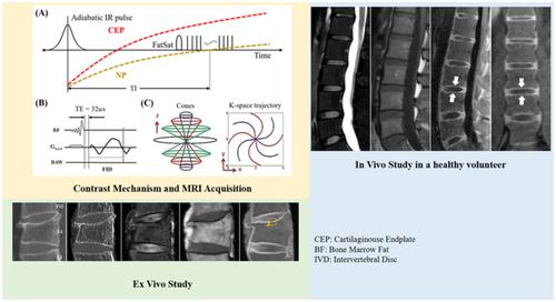
Ultrashort echo time (UTE) sequences can image tissues with transverse T 2/T 2* relaxations too short to be efficiently observed on routine clinical MRI sequences, such as the vertebral body cartilaginous endplate (CEP). Here, we describe a 3D adiabatic inversion-recovery-prepared fat-saturated ultrashort echo time (3D IR-FS-UTE) sequence to highlight the CEP of vertebral bodies in comparison to the intervertebral disc (IVD) and bone marrow fat (BF) at 3 T. The IR-FS-UTE sequence used a 3D UTE sequence combined with an adiabatic IR preparation pulse centered in the middle of the water and fat peaks, while a fat saturation module was used to suppress the signal from fat. A slab-selective half pulse was used for signal excitation, and a 3D center-out cones trajectory was used for more efficient data sampling. The 3D IR-FS-UTE sequence was applied to an ex vivo human spine sample, as well as the spines of six healthy volunteers and of three patients with back pain. Bright continuous lines representing signal from CEP were found in healthy IVDs. The measured contrast-to-noise ratio was 18.5 ± 4.9 between the CEP and BF, and 20.3 ± 4.15 between the CEP and IVD for the six volunteers. Abnormal IVDs showed CEP discontinuity or irregularity in the sample and patient studies. In conclusion, the proposed 3D IR-FS-UTE sequence is feasible for imaging the vertebral body's CEP in vivo with high contrast.
中文翻译:

使用 3D 绝热反转恢复准备脂肪饱和超短回波时间 (3D IR-FS-UTE) 序列的高对比度软骨终板成像
超短回波时间 (UTE) 序列可以对具有横向T 2 / T 2的组织进行成像* 松弛太短,无法在常规临床 MRI 序列(如椎体软骨终板 (CEP))上有效观察到。在这里,我们描述了一个 3D 绝热反转恢复准备的脂肪饱和超短回波时间 (3D IR-FS-UTE) 序列,以突出椎体的 CEP 与椎间盘 (IVD) 和骨髓脂肪 (BF) 相比在 3 T。IR-FS-UTE 序列使用 3D UTE 序列与以水和脂肪峰中间为中心的绝热 IR 准备脉冲相结合,而脂肪饱和模块用于抑制来自脂肪的信号。平板选择性半脉冲用于信号激励,3D 中心外锥轨迹用于更有效的数据采样。将 3D IR-FS-UTE 序列应用于离体人体脊柱样本,以及六名健康志愿者和三名背痛患者的脊椎。在健康的体外诊断设备中发现了代表来自 CEP 的信号的明亮连续线。六名志愿者在 CEP 和 BF 之间测量的对比度噪声比为 18.5 ± 4.9,在 CEP 和 IVD 之间为 20.3 ± 4.15。异常 IVD 在样本和患者研究中显示 CEP 不连续或不规则。总之,所提出的 3D IR-FS-UTE 序列对于体内高对比度的椎体 CEP 成像是可行的。异常 IVD 在样本和患者研究中显示 CEP 不连续或不规则。总之,所提出的 3D IR-FS-UTE 序列对于体内高对比度的椎体 CEP 成像是可行的。异常 IVD 在样本和患者研究中显示 CEP 不连续或不规则。总之,所提出的 3D IR-FS-UTE 序列对于体内高对比度的椎体 CEP 成像是可行的。
更新日期:2021-09-08
中文翻译:
使用 3D 绝热反转恢复准备脂肪饱和超短回波时间 (3D IR-FS-UTE) 序列的高对比度软骨终板成像
超短回波时间 (UTE) 序列可以对具有横向T 2 / T 2的组织进行成像* 松弛太短,无法在常规临床 MRI 序列(如椎体软骨终板 (CEP))上有效观察到。在这里,我们描述了一个 3D 绝热反转恢复准备的脂肪饱和超短回波时间 (3D IR-FS-UTE) 序列,以突出椎体的 CEP 与椎间盘 (IVD) 和骨髓脂肪 (BF) 相比在 3 T。IR-FS-UTE 序列使用 3D UTE 序列与以水和脂肪峰中间为中心的绝热 IR 准备脉冲相结合,而脂肪饱和模块用于抑制来自脂肪的信号。平板选择性半脉冲用于信号激励,3D 中心外锥轨迹用于更有效的数据采样。将 3D IR-FS-UTE 序列应用于离体人体脊柱样本,以及六名健康志愿者和三名背痛患者的脊椎。在健康的体外诊断设备中发现了代表来自 CEP 的信号的明亮连续线。六名志愿者在 CEP 和 BF 之间测量的对比度噪声比为 18.5 ± 4.9,在 CEP 和 IVD 之间为 20.3 ± 4.15。异常 IVD 在样本和患者研究中显示 CEP 不连续或不规则。总之,所提出的 3D IR-FS-UTE 序列对于体内高对比度的椎体 CEP 成像是可行的。异常 IVD 在样本和患者研究中显示 CEP 不连续或不规则。总之,所提出的 3D IR-FS-UTE 序列对于体内高对比度的椎体 CEP 成像是可行的。异常 IVD 在样本和患者研究中显示 CEP 不连续或不规则。总之,所提出的 3D IR-FS-UTE 序列对于体内高对比度的椎体 CEP 成像是可行的。


 京公网安备 11010802027423号