Our official English website, www.x-mol.net, welcomes your feedback! (Note: you will need to create a separate account there.)
Multimodal Imaging with NanoGd Reveals Spatiotemporal Features of Neuroinflammation after Experimental Stroke
Advanced Science ( IF 15.1 ) Pub Date : 2021-07-01 , DOI: 10.1002/advs.202101433 Violaine Hubert 1 , Ines Hristovska 2 , Szilvia Karpati 3 , Sarah Benkeder 2 , Arindam Dey 4 , Chloé Dumot 1 , Camille Amaz 5 , Naura Chounlamountri 2 , Chantal Watrin 2 , Jean-Christophe Comte 6 , Fabien Chauveau 7 , Emmanuel Brun 8 , Patrice Marche 4 , Fréderic Lerouge 3 , Stéphane Parola 3 , Yves Berthezène 9 , Thomas Vorup-Jensen 10 , Olivier Pascual 2 , Marlène Wiart 1
Advanced Science ( IF 15.1 ) Pub Date : 2021-07-01 , DOI: 10.1002/advs.202101433 Violaine Hubert 1 , Ines Hristovska 2 , Szilvia Karpati 3 , Sarah Benkeder 2 , Arindam Dey 4 , Chloé Dumot 1 , Camille Amaz 5 , Naura Chounlamountri 2 , Chantal Watrin 2 , Jean-Christophe Comte 6 , Fabien Chauveau 7 , Emmanuel Brun 8 , Patrice Marche 4 , Fréderic Lerouge 3 , Stéphane Parola 3 , Yves Berthezène 9 , Thomas Vorup-Jensen 10 , Olivier Pascual 2 , Marlène Wiart 1
Affiliation
|
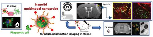
The purpose of this study is to propose and validate a preclinical in vivo magnetic resonance imaging (MRI) tool to monitor neuroinflammation following ischemic stroke, based on injection of a novel multimodal nanoprobe, NanoGd, specifically designed for internalization by phagocytic cells. First, it is verified that NanoGd is efficiently internalized by microglia in vitro. In vivo MRI coupled with intravenous injection of NanoGd in a permanent middle cerebral artery occlusion mouse model results in hypointense signals in the ischemic lesion. In these mice, longitudinal two-photon intravital microscopy shows NanoGd internalization by activated CX3CR1-GFP/+ cells. Ex vivo analysis, including phase contrast imaging with synchrotron X-ray, histochemistry, and transmission electron microscopy corroborate NanoGd accumulation within the ischemic lesion and uptake by immune phagocytic cells. Taken together, these results confirm the potential of NanoGd-enhanced MRI as an imaging biomarker of neuroinflammation at the subacute stage of ischemic stroke. As far as it is known, this work is the first to decipher the working mechanism of MR signals induced by a nanoparticle passively targeted at phagocytic cells by performing intravital microscopy back-to-back with MRI. Furthermore, using a gadolinium-based rather than an iron-based contrast agent raises future perspectives for the development of molecular imaging with emerging computed tomography technologies.
中文翻译:

NanoGd 多模态成像揭示实验性中风后神经炎症的时空特征
本研究的目的是提出并验证一种临床前体内磁共振成像 (MRI) 工具,用于监测缺血性中风后的神经炎症,该工具基于注射一种新型多模式纳米探针 NanoGd,该探针专为吞噬细胞内化而设计。首先,在体外验证了NanoGd能够被小胶质细胞有效内化。在永久性大脑中动脉闭塞小鼠模型中,体内 MRI 结合静脉注射 NanoGd 会导致缺血性病变中出现低信号。在这些小鼠中,纵向双光子活体显微镜显示 NanoGd 被激活的 CX3CR1-GFP/+ 细胞内化。离体分析,包括同步加速器 X 射线相差成像、组织化学和透射电子显微镜,证实了 NanoGd 在缺血性病变内的积累和免疫吞噬细胞的摄取。总而言之,这些结果证实了 NanoGd 增强 MRI 作为缺血性中风亚急性阶段神经炎症成像生物标志物的潜力。据了解,这项工作首次通过与MRI背对背进行活体显微镜检查,破译了纳米颗粒被动靶向吞噬细胞诱导MR信号的工作机制。此外,使用钆基造影剂而不是铁基造影剂为新兴计算机断层扫描技术的分子成像发展提出了未来前景。
更新日期:2021-09-09
中文翻译:
NanoGd 多模态成像揭示实验性中风后神经炎症的时空特征
本研究的目的是提出并验证一种临床前体内磁共振成像 (MRI) 工具,用于监测缺血性中风后的神经炎症,该工具基于注射一种新型多模式纳米探针 NanoGd,该探针专为吞噬细胞内化而设计。首先,在体外验证了NanoGd能够被小胶质细胞有效内化。在永久性大脑中动脉闭塞小鼠模型中,体内 MRI 结合静脉注射 NanoGd 会导致缺血性病变中出现低信号。在这些小鼠中,纵向双光子活体显微镜显示 NanoGd 被激活的 CX3CR1-GFP/+ 细胞内化。离体分析,包括同步加速器 X 射线相差成像、组织化学和透射电子显微镜,证实了 NanoGd 在缺血性病变内的积累和免疫吞噬细胞的摄取。总而言之,这些结果证实了 NanoGd 增强 MRI 作为缺血性中风亚急性阶段神经炎症成像生物标志物的潜力。据了解,这项工作首次通过与MRI背对背进行活体显微镜检查,破译了纳米颗粒被动靶向吞噬细胞诱导MR信号的工作机制。此外,使用钆基造影剂而不是铁基造影剂为新兴计算机断层扫描技术的分子成像发展提出了未来前景。


 京公网安备 11010802027423号