当前位置:
X-MOL 学术
›
Lett. Appl. Microbiol.
›
论文详情
Our official English website, www.x-mol.net, welcomes your
feedback! (Note: you will need to create a separate account there.)
Biofilm prevalence and microbial characterisation in chronic wounds in a Sri Lankan cohort
Letters in Applied Microbiology ( IF 2.0 ) Pub Date : 2021-06-29 , DOI: 10.1111/lam.13532 A Dilhari 1, 2 , M Weerasekera 1 , C Gunasekara 1 , S Pathirage 3 , N Fernando 1 , D Weerasekara 4 , A J McBain 1, 5
Letters in Applied Microbiology ( IF 2.0 ) Pub Date : 2021-06-29 , DOI: 10.1111/lam.13532 A Dilhari 1, 2 , M Weerasekera 1 , C Gunasekara 1 , S Pathirage 3 , N Fernando 1 , D Weerasekara 4 , A J McBain 1, 5
Affiliation
|
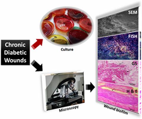
Biofilms have been associated with chronic wound infections in diabetic patients. The study assessed the occurrence of biofilms in chronic diabetic wounds (CDWs) in a Sri Lankan cohort. Tissue specimens collected during surgical debridement were analysed by quantitative differential viable counting, scanning electron microscopy (SEM), fluorescence insitu hybridization (FISH) and light microscopy with Gram and Haematoxylin-Eosin staining. All specimens harboured >5·0 log10 CFU per g bacteria and 2–9 distinct species per specimen were recovered from twenty wounds by culture. The most frequently isolated bacterium was Pseudomonas spp. (12/20;60%). Strict anaerobes were isolated from 10/20 specimens. Gram and Haematoxylin-Eosin staining showed aggregated micro-colonies, embedded in the wound tissue bed (20/20) but the exopolymer matrix was not visible in all samples (13/20). Fluorescence microscopy using a eubacteria-specific FISH probe indicated the presence of bacterial aggregates within the deep layers of the wound tissues (20/20). SEM revealed the presumptive architecture of matrix-embedded microbial clusters (20/20). The approximate diameter of bacterial aggregates in tissues ranged between 12 and 400 µm. Bacterial infiltration into the internal portions of the tissues was apparent using FISH, Gram, and Haematoxylin-Eosin staining. All CDWs carried biofilm-specific morphological features. FISH was more specific than SEM and indicated the presence of microcolonies within deeper tissues.
中文翻译:

斯里兰卡队列慢性伤口的生物膜流行率和微生物特征
生物膜与糖尿病患者的慢性伤口感染有关。该研究评估了斯里兰卡队列中慢性糖尿病伤口 (CDW) 中生物膜的发生情况。在手术清创期间收集的组织标本通过定量差分活菌计数、扫描电子显微镜 (SEM)、荧光原位杂交 (FISH) 和革兰氏和苏木精-伊红染色的光学显微镜进行分析。所有标本每克细菌含有 >5·0 log 10 CFU,每个标本通过培养从 20 个伤口中回收了 2-9 个不同的物种。最常分离的细菌是假单胞菌属 (12/20;60%)。从 10/20 样本中分离出严格的厌氧菌。革兰氏和苏木精-伊红染色显示聚集的微菌落,嵌入伤口组织床 (20/20),但外聚体基质在所有样品中均不可见 (13/20)。使用真细菌特异性 FISH 探针的荧光显微镜显示伤口组织深层内存在细菌聚集体 (20/20)。SEM 揭示了基质嵌入微生物簇的假定结构 (20/20)。组织中细菌聚集体的近似直径介于 12 和 400 µ之间 米。使用 FISH、革兰氏和苏木精-伊红染色可以明显看出细菌浸润到组织内部。所有 CDW 都具有生物膜特定的形态特征。FISH 比 SEM 更具特异性,表明在更深的组织中存在微菌落。
更新日期:2021-06-29
中文翻译:
斯里兰卡队列慢性伤口的生物膜流行率和微生物特征
生物膜与糖尿病患者的慢性伤口感染有关。该研究评估了斯里兰卡队列中慢性糖尿病伤口 (CDW) 中生物膜的发生情况。在手术清创期间收集的组织标本通过定量差分活菌计数、扫描电子显微镜 (SEM)、荧光原位杂交 (FISH) 和革兰氏和苏木精-伊红染色的光学显微镜进行分析。所有标本每克细菌含有 >5·0 log 10 CFU,每个标本通过培养从 20 个伤口中回收了 2-9 个不同的物种。最常分离的细菌是假单胞菌属 (12/20;60%)。从 10/20 样本中分离出严格的厌氧菌。革兰氏和苏木精-伊红染色显示聚集的微菌落,嵌入伤口组织床 (20/20),但外聚体基质在所有样品中均不可见 (13/20)。使用真细菌特异性 FISH 探针的荧光显微镜显示伤口组织深层内存在细菌聚集体 (20/20)。SEM 揭示了基质嵌入微生物簇的假定结构 (20/20)。组织中细菌聚集体的近似直径介于 12 和 400 µ之间 米。使用 FISH、革兰氏和苏木精-伊红染色可以明显看出细菌浸润到组织内部。所有 CDW 都具有生物膜特定的形态特征。FISH 比 SEM 更具特异性,表明在更深的组织中存在微菌落。


 京公网安备 11010802027423号