当前位置:
X-MOL 学术
›
Immunology
›
论文详情
Our official English website, www.x-mol.net, welcomes your
feedback! (Note: you will need to create a separate account there.)
PD-1-induced proliferating T cells exhibit a distinct transcriptional signature
Immunology ( IF 4.9 ) Pub Date : 2021-06-23 , DOI: 10.1111/imm.13388 Marianne Strazza 1 , Shoiab Bukhari 1 , Anna S Tocheva 1 , Adam Mor 1
Immunology ( IF 4.9 ) Pub Date : 2021-06-23 , DOI: 10.1111/imm.13388 Marianne Strazza 1 , Shoiab Bukhari 1 , Anna S Tocheva 1 , Adam Mor 1
Affiliation
|
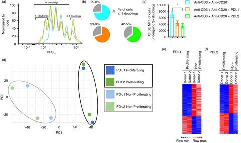
Ligation of the inhibitory receptor PD-1 on T cells results in the inhibition of numerous cellular functions. Despite the overtly inhibitory outcome of PD-1 signalling, there are additionally a collection of functions that are activated. We have observed that CD4+ T cells stimulated through the T-cell receptor and PD-1 primarily do not proliferate; however, there is a population of cells that proliferates more than T-cell receptor stimulation alone. These highly proliferating cells could potentially be associated with PD-1-blockade unresponsiveness in patients. In this study, we have performed RNA sequencing and found that following PD-1 ligation proliferating and non-proliferating T cells have distinct transcriptional signatures. Remarkably, the proliferating cells showed an enrichment of genes associated with an activated state despite PD-1 signalling. Additionally, circulating follicular helper T cells were significantly more prevalent in the non-proliferating population, demonstrated by enrichment of the associated genes CXCR5, CCR7, TCF7, BCL6 and PRDM1 and validated at the protein level. Translationally, we also show that there are more follicular helper T cells in patients that respond favourably to PD-1 blockade. Overall, the presence of transcriptionally and functionally distinct T cell populations responsive to PD-1 ligation may provide insights into the clinical differences observed following therapeutic PD-1 blockade.
中文翻译:

PD-1诱导的增殖T细胞表现出独特的转录特征
T 细胞上的抑制性受体 PD-1 的连接会导致多种细胞功能的抑制。尽管 PD-1 信号传导具有明显的抑制作用,但还有一系列功能被激活。我们观察到,通过 T 细胞受体和 PD-1 刺激的 CD4 + T 细胞主要不增殖;然而,有一群细胞的增殖速度比单独刺激 T 细胞受体要快。这些高度增殖的细胞可能与患者对 PD-1 阻断无反应有关。在这项研究中,我们进行了 RNA 测序,发现在 PD-1 连接后,增殖和非增殖 T 细胞具有不同的转录特征。值得注意的是,尽管存在 PD-1 信号传导,但增殖细胞显示出与激活状态相关的基因富集。此外,循环滤泡辅助 T 细胞在非增殖群体中明显更为普遍,相关基因CXCR5 、 CCR7 、 TCF7 、 BCL6和PRDM1的富集证明了这一点,并在蛋白质水平上进行了验证。从转化角度来看,我们还表明,患者体内有更多的滤泡辅助 T 细胞,对 PD-1 阻断有良好的反应。总体而言,对 PD-1 连接有反应的转录和功能不同的 T 细胞群的存在可能有助于了解治疗性 PD-1 阻断后观察到的临床差异。
更新日期:2021-06-23
中文翻译:
PD-1诱导的增殖T细胞表现出独特的转录特征
T 细胞上的抑制性受体 PD-1 的连接会导致多种细胞功能的抑制。尽管 PD-1 信号传导具有明显的抑制作用,但还有一系列功能被激活。我们观察到,通过 T 细胞受体和 PD-1 刺激的 CD4 + T 细胞主要不增殖;然而,有一群细胞的增殖速度比单独刺激 T 细胞受体要快。这些高度增殖的细胞可能与患者对 PD-1 阻断无反应有关。在这项研究中,我们进行了 RNA 测序,发现在 PD-1 连接后,增殖和非增殖 T 细胞具有不同的转录特征。值得注意的是,尽管存在 PD-1 信号传导,但增殖细胞显示出与激活状态相关的基因富集。此外,循环滤泡辅助 T 细胞在非增殖群体中明显更为普遍,相关基因CXCR5 、 CCR7 、 TCF7 、 BCL6和PRDM1的富集证明了这一点,并在蛋白质水平上进行了验证。从转化角度来看,我们还表明,患者体内有更多的滤泡辅助 T 细胞,对 PD-1 阻断有良好的反应。总体而言,对 PD-1 连接有反应的转录和功能不同的 T 细胞群的存在可能有助于了解治疗性 PD-1 阻断后观察到的临床差异。


 京公网安备 11010802027423号