Chemical Engineering Journal ( IF 13.3 ) Pub Date : 2021-06-24 , DOI: 10.1016/j.cej.2021.130961 Huan Sun , Chenxi Zhang , Boqing Zhang , Ping Song , Xiujuan Xu , Xingyu Gui , Xinyue Chen , Gonggong Lu , Xiang Li , Jie Liang , Jianxun Sun , Qing Jiang , Changchun Zhou , Yujiang Fan , Xuedong Zhou , Xingdong Zhang
|
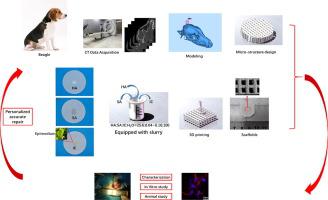
The purpose of this research was to repair critical size defects of a skull. First, the CT data on the Beagle's skull were used to reconstruct an implant model. Then two Ф15 mm critical bone defects on the left and right sides of the skull were drilled. Hydroxyapatite, sodium alginate and icariin were adopted to formulate the printing inks according to their proportions. Calcium phosphate scaffolds with accuracy porosity were 3D printed by an extrusion printer. Icariin served as a controlled release of osteogenic drugs for bone regeneration. The physical and chemical properties of the 3D printed scaffolds were summarized, and in vitro and in vivo tests were conducted to assess the biocompatibility and the bone repair ability of the scaffolds. According to the mechanical results, the 3D printed calcium phosphate scaffolds show improved mechanical strength, and the Young's modulus was about 4.23 MPa, while the compressive strength was 9.37 MPa. The drug release test results showed that the slowly controlled release of osteogenic drugs behavior by the 3D printed calcium phosphate scaffolds continuously promoted new bone formation. The in vitro biological assessment results suggested that the 3D printed calcium phosphate scaffolds had good biocompatibility, with no obvious cytotoxicity observed. An animal model, aimed at achieving personalized accurate repair of the critical size defects on the skull, indicated that the 3D printed calcium phosphate scaffolds with controlled release of osteogenic drugs had great potential for regeneration of critical bone defects.
中文翻译:
3D 打印磷酸钙支架,控制释放成骨药物用于骨再生
这项研究的目的是修复颅骨的关键尺寸缺陷。首先,Beagle 颅骨的 CT 数据用于重建植入模型。然后在颅骨左右两侧分别钻出两个Ф15 mm的严重骨缺损。采用羟基磷灰石、海藻酸钠和淫羊藿苷按比例配制印刷油墨。具有精确孔隙率的磷酸钙支架由挤出打印机 3D 打印。淫羊藿苷作为成骨药物的受控释放用于骨再生。总结了3D打印支架的理化性质,并进行了体外和体内试验,评估了支架的生物相容性和骨修复能力。根据力学结果,3D打印的磷酸钙支架显示出更高的机械强度,杨氏模量约为4.23 MPa,而抗压强度为9.37 MPa。药物释放测试结果表明,3D打印磷酸钙支架缓慢控制释放成骨药物行为,持续促进新骨形成。体外生物学评估结果表明,3D打印磷酸钙支架具有良好的生物相容性,未观察到明显的细胞毒性。一个旨在实现颅骨临界尺寸缺损个性化精确修复的动物模型表明,具有控释成骨药物的3D打印磷酸钙支架在再生关键骨缺损方面具有巨大潜力。而抗压强度为 9.37 MPa。药物释放测试结果表明,3D打印磷酸钙支架缓慢控制释放成骨药物行为,持续促进新骨形成。体外生物学评估结果表明,3D打印磷酸钙支架具有良好的生物相容性,未观察到明显的细胞毒性。一个旨在实现颅骨临界尺寸缺损个性化精确修复的动物模型表明,具有控释成骨药物的3D打印磷酸钙支架在再生关键骨缺损方面具有巨大潜力。而抗压强度为 9.37 MPa。药物释放测试结果表明,3D打印磷酸钙支架缓慢控制释放成骨药物行为,持续促进新骨形成。体外生物学评估结果表明,3D打印磷酸钙支架具有良好的生物相容性,未观察到明显的细胞毒性。一个旨在实现颅骨临界尺寸缺损个性化精确修复的动物模型表明,具有控释成骨药物的3D打印磷酸钙支架在再生关键骨缺损方面具有巨大潜力。药物释放测试结果表明,3D打印磷酸钙支架缓慢控制释放成骨药物行为,持续促进新骨形成。体外生物学评估结果表明,3D打印磷酸钙支架具有良好的生物相容性,未观察到明显的细胞毒性。一个旨在实现颅骨临界尺寸缺损个性化精确修复的动物模型表明,具有控释成骨药物的3D打印磷酸钙支架在再生关键骨缺损方面具有巨大潜力。药物释放测试结果表明,3D打印磷酸钙支架缓慢控制释放成骨药物行为,持续促进新骨形成。体外生物学评估结果表明,3D打印磷酸钙支架具有良好的生物相容性,未观察到明显的细胞毒性。一个旨在实现颅骨临界尺寸缺损个性化精确修复的动物模型表明,具有控释成骨药物的3D打印磷酸钙支架在再生关键骨缺损方面具有巨大潜力。


 京公网安备 11010802027423号