当前位置:
X-MOL 学术
›
NMR Biomed.
›
论文详情
Our official English website, www.x-mol.net, welcomes your feedback! (Note: you will need to create a separate account there.)
Deuterium MRSI characterizations of glucose metabolism in orthotopic pancreatic cancer mouse models
NMR in Biomedicine ( IF 2.9 ) Pub Date : 2021-06-16 , DOI: 10.1002/nbm.4569 Stefan Markovic 1 , Tangi Roussel 2 , Lilach Agemy 3 , Keren Sasson 3 , Dina Preise 4 , Avigdor Scherz 3 , Lucio Frydman 1
NMR in Biomedicine ( IF 2.9 ) Pub Date : 2021-06-16 , DOI: 10.1002/nbm.4569 Stefan Markovic 1 , Tangi Roussel 2 , Lilach Agemy 3 , Keren Sasson 3 , Dina Preise 4 , Avigdor Scherz 3 , Lucio Frydman 1
Affiliation
|
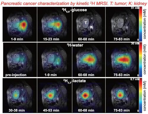
Detecting and mapping metabolism in tissues represents a major step in detecting, characterizing, treating and understanding cancers. Recently introduced deuterium metabolic imaging techniques could offer a noninvasive route for the metabolic imaging of animals and humans, based on using 2H magnetic resonance spectroscopic imaging (MRSI) to detect the uptake of deuterated glucose and the fate of its metabolic products. In this study, 2H6,6′-glucose was administered to mice cohorts that had been orthotopically implanted with two different models of pancreatic ductal adenocarcinoma (PDAC), involving PAN-02 and KPC cell lines. As the tumors grew, 2H6,6′-glucose was administered as bolii into the animals' tail veins, and 2H MRSI images were recorded at 15.2 T. 2D phase-encoded chemical shift imaging experiments could detect a signal from this deuterated glucose immediately after the bolus injection for both the PDAC models, reaching a maximum in the animals' tumors ~ 20 min following administration, and nearly total decay after ~ 40 min. The main metabolic reporter of the cancers was the 2H3,3′-lactate signal, which MRSI could detect and localize on the tumors when these were 5 mm or more in diameter. Lactate production time traces varied slightly with the animal and tumor model, but in general lactate peaked at times of 60 min or longer following injection, reaching concentrations that were ~ 10-fold lower than those of the initial glucose injection. This 2H3,3′-lactate signal was only visible inside the tumors. 2H-water could also be detected as deuterated glucose's metabolic product, increasing throughout the entire time course of the experiment from its ≈10 mM natural abundance background. This water resonance could be imaged throughout the entire abdomen of the animals, including an enhanced presence in the tumor, but also in other organs like the kidney and bladder. These results suggest that deuterium MRSI may serve as a robust, minimally invasive tool for the monitoring of metabolic activity in pancreatic tumors, capable of undergoing clinical translation and supporting decisions concerning treatment strategies. Comparisons with in vivo metabolic MRI experiments that have been carried out in other animal models are presented and their differences/similarities are discussed.
中文翻译:

原位胰腺癌小鼠模型中葡萄糖代谢的氘 MRSI 表征
检测和绘制组织中的新陈代谢是检测、表征、治疗和了解癌症的重要步骤。最近引入的氘代谢成像技术可以为动物和人类的代谢成像提供一种无创途径,基于使用2 H 磁共振波谱成像 (MRSI) 检测氘代葡萄糖的摄取及其代谢产物的命运。在这项研究中,将2 H 6,6' -葡萄糖施用于已原位植入两种不同模型的胰腺导管腺癌 (PDAC) 的小鼠队列,包括 PAN-02 和 KPC 细胞系。随着肿瘤的生长,2 H 6,6′-葡萄糖以bolii形式注入动物的尾静脉,并在15.2 T记录2 H MRSI图像。2D相位编码化学位移成像实验可以在两种PDAC模型的推注后立即检测到来自这种氘化葡萄糖的信号,在给药后约 20 分钟在动物的肿瘤中达到最大值,约 40 分钟后几乎完全衰减。癌症的主要代谢报告基因是2 H 3,3'-乳酸信号,当肿瘤直径为 5 mm 或更大时,MRSI 可以检测并定位在肿瘤上。乳酸产生时间轨迹随动物和肿瘤模型的不同而略有不同,但一般来说,乳酸在注射后 60 分钟或更长时间达到峰值,达到比初始葡萄糖注射低约 10 倍的浓度。这种2 H 3,3' -乳酸信号仅在肿瘤内部可见。2H-水也可以被检测为氘代葡萄糖的代谢产物,在整个实验过程中从其 ≈10 mM 的自然丰度背景增加。这种水共振可以在动物的整个腹部成像,包括在肿瘤中增强的存在,但也可以在肾脏和膀胱等其他器官中。这些结果表明,氘 MRSI 可作为一种强大的微创工具,用于监测胰腺肿瘤的代谢活动,能够进行临床转化并支持有关治疗策略的决策。与已在其他动物模型中进行的体内代谢 MRI 实验进行了比较,并讨论了它们的差异/相似之处。
更新日期:2021-08-11
中文翻译:
原位胰腺癌小鼠模型中葡萄糖代谢的氘 MRSI 表征
检测和绘制组织中的新陈代谢是检测、表征、治疗和了解癌症的重要步骤。最近引入的氘代谢成像技术可以为动物和人类的代谢成像提供一种无创途径,基于使用2 H 磁共振波谱成像 (MRSI) 检测氘代葡萄糖的摄取及其代谢产物的命运。在这项研究中,将2 H 6,6' -葡萄糖施用于已原位植入两种不同模型的胰腺导管腺癌 (PDAC) 的小鼠队列,包括 PAN-02 和 KPC 细胞系。随着肿瘤的生长,2 H 6,6′-葡萄糖以bolii形式注入动物的尾静脉,并在15.2 T记录2 H MRSI图像。2D相位编码化学位移成像实验可以在两种PDAC模型的推注后立即检测到来自这种氘化葡萄糖的信号,在给药后约 20 分钟在动物的肿瘤中达到最大值,约 40 分钟后几乎完全衰减。癌症的主要代谢报告基因是2 H 3,3'-乳酸信号,当肿瘤直径为 5 mm 或更大时,MRSI 可以检测并定位在肿瘤上。乳酸产生时间轨迹随动物和肿瘤模型的不同而略有不同,但一般来说,乳酸在注射后 60 分钟或更长时间达到峰值,达到比初始葡萄糖注射低约 10 倍的浓度。这种2 H 3,3' -乳酸信号仅在肿瘤内部可见。2H-水也可以被检测为氘代葡萄糖的代谢产物,在整个实验过程中从其 ≈10 mM 的自然丰度背景增加。这种水共振可以在动物的整个腹部成像,包括在肿瘤中增强的存在,但也可以在肾脏和膀胱等其他器官中。这些结果表明,氘 MRSI 可作为一种强大的微创工具,用于监测胰腺肿瘤的代谢活动,能够进行临床转化并支持有关治疗策略的决策。与已在其他动物模型中进行的体内代谢 MRI 实验进行了比较,并讨论了它们的差异/相似之处。


 京公网安备 11010802027423号