Annals of Anatomy ( IF 2.0 ) Pub Date : 2021-06-16 , DOI: 10.1016/j.aanat.2021.151785 Cesar Rios-Navarro 1 , Nuria Daghbouche-Rubio 1 , Jose Gavara 1 , Elena de Dios 2 , Nerea Perez 1 , Jose M Vila 3 , Francisco J Chorro 4 , Amparo Ruiz-Sauri 5 , Vicente Bodi 4
|
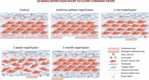
Background
Coronary arteries supply oxygen and nutrients to the heart. We evaluated the dynamics of microscopic damage throughout the ischemia-reperfusion process in the wall of coronary arteries following myocardial infarction (MI).
Methods
In a swine model of reperfused MI, animals were divided into one control and four MI groups: 90-min ischemia without reperfusion, or followed by one minute, one week or one month reperfusion. Left anterior descending (LAD; infarct-related artery) and control right coronary arteries (RCA) were isolated. Taking the balloon inflation region as a reference, we isolated the proximal and distal LAD areas, performing histological staining and immunohistochemistry.
Results
Although mild changes in tunica intima were observed during ischemia, an almost complete absence of endothelium, and abnormal breaks in the internal elastic layer were found post-revascularization. In tunica media, increased thickness was observed soon after reperfusion, whereas larger thickness, disorganized muscle cell distribution and edema were found one week after reperfusion. This damage was more pronounced in distal rather than proximal LAD, whereas no changes were detected in RCA. In the tunica adventitia, vasa vasorum density decayed during ischemia in both LAD regions, but was restored after one month. Leukocyte adhesion to the artery was observed post-revascularization, developing into a massive presence in the three layers one week post-reperfusion.
Conclusions
Ischemia-reperfusion can itself induce damage in the wall of the epicardial coronary artery, becoming more pronounced in the region distal to balloon inflation. Exploring these abnormalities will provide insight into the pathophysiology of coronary circulation and MI.
中文翻译:
冠状动脉缺血再灌注损伤:再灌注心肌梗死后的综合显微研究
背景
冠状动脉为心脏提供氧气和营养。我们评估了心肌梗死 (MI) 后冠状动脉壁缺血再灌注过程中微观损伤的动态。
方法
在再灌注心肌梗死的猪模型中,动物被分为一个对照组和四个心肌梗死组:90 分钟缺血无再灌注,或随后再灌注 1 分钟、1 周或 1 个月。分离左前降支(LAD;梗塞相关动脉)和对照右冠状动脉(RCA)。以球囊膨胀区域为参考,我们分离了近端和远端 LAD 区域,进行组织学染色和免疫组织化学。
结果
尽管在缺血期间观察到内膜的轻微变化,但在血运重建后发现内皮几乎完全缺失和内部弹性层异常断裂。在中膜中,再灌注后很快观察到厚度增加,而再灌注后一周发现厚度增加、肌细胞分布紊乱和水肿。这种损伤在远端而不是近端 LAD 中更明显,而在 RCA 中没有检测到变化。在外膜中,两个 LAD 区域的血管滋养管密度在缺血期间均下降,但在 1 个月后恢复。血运重建后观察到白细胞粘附于动脉,再灌注后一周内在三层中大量存在。
结论
缺血再灌注本身可引起心外膜冠状动脉壁的损伤,在球囊膨胀远端的区域变得更加明显。探索这些异常将有助于深入了解冠状动脉循环和 MI 的病理生理学。


 京公网安备 11010802027423号