当前位置:
X-MOL 学术
›
NMR Biomed.
›
论文详情
Our official English website, www.x-mol.net, welcomes your
feedback! (Note: you will need to create a separate account there.)
Paradoxical paramagnetic calcifications in the globus pallidus: An ex vivo MR investigation and histological validation study
NMR in Biomedicine ( IF 2.7 ) Pub Date : 2021-06-15 , DOI: 10.1002/nbm.4571 Jinhee Jang 1 , Yoonho Nam 1, 2 , Sung Won Jung 3 , Tae-Ryong Riew 3, 4 , Sang Hyun Kim 3 , In-Beom Kim 3, 4
NMR in Biomedicine ( IF 2.7 ) Pub Date : 2021-06-15 , DOI: 10.1002/nbm.4571 Jinhee Jang 1 , Yoonho Nam 1, 2 , Sung Won Jung 3 , Tae-Ryong Riew 3, 4 , Sang Hyun Kim 3 , In-Beom Kim 3, 4
Affiliation
|
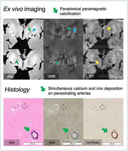
MR images based on phase contrast images have gained clinical interest as an in vivo tool for assessing anatomical and histological findings. The globus pallidus is an area of major iron metabolism and storage in the brain tissue. Calcium, another important metal in the body, is frequently deposited in the globus pallidus as well. Recently, we observed dense paramagnetic deposition with paradoxical calcifications in the globus pallidus and putamen. In this work, we explore detailed MR findings on these structures, and the histological source of the related findings using ex vivo CT and MR images. Ex vivo MR was obtained with a maximum 100 μm3 isotropic resolution using a 15.2 T MR system. 3D gradient echo images and quantitative susceptibility mapping were used because of their good sensitivity to metallic deposition, high signal-to-noise ratio, and excellent contrast to iron and calcium. We found dense paramagnetic deposition along the perforating arteries in the globus pallidus. This paramagnetic deposition was hyperdense on ex vivo CT scans. Histological studies confirmed this finding, and simultaneous deposition of iron and calcium, although more iron dominant, was observed along the vessel walls of the globus pallidus. This was an exclusive finding for the penetrating arteries of the globus pallidus. Thus, our results suggest that several strong and paradoxical paramagnetic sources at the globus pallidus can be associated with vascular degeneration.
中文翻译:

苍白球中反常的顺磁性钙化:离体 MR 调查和组织学验证研究
基于相位对比图像的 MR 图像作为一种用于评估解剖和组织学发现的体内工具已获得临床兴趣。苍白球是脑组织中主要的铁代谢和储存区域。钙是体内另一种重要的金属,也经常沉积在苍白球中。最近,我们观察到在苍白球和壳核中具有反常钙化的致密顺磁沉积。在这项工作中,我们探索了这些结构的详细 MR 发现,以及使用离体 CT 和 MR 图像相关发现的组织学来源。离体 MR 最大 100 μm 3使用 15.2 T MR 系统的各向同性分辨率。使用 3D 梯度回波图像和定量磁化率映射是因为它们对金属沉积具有良好的敏感性、高信噪比以及与铁和钙的出色对比度。我们在苍白球的穿支动脉上发现了密集的顺磁沉积。这种顺磁沉积在离体 CT 扫描中是高密度的。组织学研究证实了这一发现,并且沿着苍白球的血管壁观察到铁和钙的同时沉积,尽管铁占主导地位。这是对苍白球穿透动脉的独家发现。因此,我们的研究结果表明,苍白球处的几个强而矛盾的顺磁源可能与血管退化有关。
更新日期:2021-06-15
中文翻译:
苍白球中反常的顺磁性钙化:离体 MR 调查和组织学验证研究
基于相位对比图像的 MR 图像作为一种用于评估解剖和组织学发现的体内工具已获得临床兴趣。苍白球是脑组织中主要的铁代谢和储存区域。钙是体内另一种重要的金属,也经常沉积在苍白球中。最近,我们观察到在苍白球和壳核中具有反常钙化的致密顺磁沉积。在这项工作中,我们探索了这些结构的详细 MR 发现,以及使用离体 CT 和 MR 图像相关发现的组织学来源。离体 MR 最大 100 μm 3使用 15.2 T MR 系统的各向同性分辨率。使用 3D 梯度回波图像和定量磁化率映射是因为它们对金属沉积具有良好的敏感性、高信噪比以及与铁和钙的出色对比度。我们在苍白球的穿支动脉上发现了密集的顺磁沉积。这种顺磁沉积在离体 CT 扫描中是高密度的。组织学研究证实了这一发现,并且沿着苍白球的血管壁观察到铁和钙的同时沉积,尽管铁占主导地位。这是对苍白球穿透动脉的独家发现。因此,我们的研究结果表明,苍白球处的几个强而矛盾的顺磁源可能与血管退化有关。


 京公网安备 11010802027423号