Journal of Materials Science: Materials in Medicine ( IF 4.2 ) Pub Date : 2021-06-14 , DOI: 10.1007/s10856-021-06548-0 Yi-Tong Yao 1 , Yue Yang 2 , Qi Ye 3 , Shan-Shan Cao 4 , Xin-Ping Zhang 4 , Ke Zhao 1 , Yutao Jian 5
|
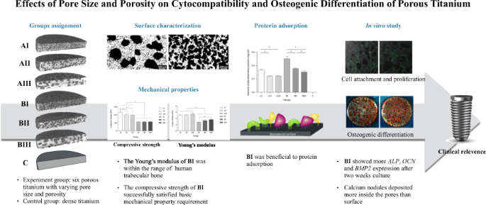
To find out the optimal porosity and pore size of porous titanium (Ti) regarding the cytocompatibility and osteogenic differentiation. Six groups of porous Ti samples with different porosities and pore sizes were fabricated by the powder metallurgy process. The microstructure and compressive mechanical properties were characterized. The cytocompatibility was examined by a series of biological tests as protein absorption with BCA assay kit, cell attachment with laser scanning confocal microscopy and vinculin expression, cell proliferation with CCK-8 assay. Cell differentiation and calcification were detected by qPCR and Alizarin Red S dying respectively. Pores distributed homogeneously throughout the porous Ti samples. The compressive test results showed that Young’s modulus ranged from 2.80 ± 0.03 GPa to 5.43 ± 0.34 GPa and the compressive strength increased from 112.4 ± 3.6 MPa to 231.1 ± 9.4 MPa. Porous Ti with high porosity (53.3 ± 1.2%) and small pore size (191.6 ± 3.7 μm) adsorbed more proteins. More MC3T3-E1 cells adhered onto dense Ti samples than onto any other porous ones already after culture and no difference was identified within the porous groups. The porous structure of porous Ti with a porosity of 53.3 ± 1.2% and an average pore size of 191.6 ± 3.7 μm facilitated cell differentiation and calcification. Small pores were not beneficial to the osteo-initiation at the very beginning. Porous Ti with a porosity of 53.3 ± 1.2% and an average pore size of 191.6 ± 3.7 μm fabricated by powder metallurgy process showed the expected mechanical property and improved osseointegration as implants in dental treatment.
中文翻译:
孔径和孔隙率对多孔钛细胞相容性和成骨分化的影响
找出多孔钛 (Ti) 在细胞相容性和成骨分化方面的最佳孔隙率和孔径。采用粉末冶金工艺制备了六组不同孔隙率和孔径的多孔钛样品。表征了微观结构和压缩力学性能。通过一系列生物学测试检查细胞相容性,如使用 BCA 检测试剂盒检测蛋白质吸收、使用激光扫描共聚焦显微镜检测细胞附着和纽蛋白表达、使用 CCK-8 检测检测细胞增殖。分别通过qPCR和茜素红S死亡检测细胞分化和钙化。孔隙均匀分布在整个多孔钛样品中。压缩试验结果表明,杨氏模量在 2.80 ± 0.03 GPa 到 5.43 ± 0 之间。34 GPa,抗压强度从112.4±3.6 MPa增加到231.1±9.4 MPa。具有高孔隙率 (53.3 ± 1.2%) 和小孔径 (191.6 ± 3.7 μm) 的多孔钛吸附更多的蛋白质。在培养后,粘附在致密 Ti 样品上的 MC3T3-E1 细胞比粘附在任何其他多孔样品上的多,并且在多孔组内没有发现差异。孔隙率为 53.3 ± 1.2%,平均孔径为 191.6 ± 3.7 μm 的多孔 Ti 的多孔结构促进了细胞分化和钙化。小孔在一开始不利于成骨。通过粉末冶金工艺制造的孔隙率为 53.3 ± 1.2% 且平均孔径为 191.6 ± 3.7 μm 的多孔钛显示出预期的机械性能和改善的骨整合,作为牙科治疗中的植入物。1 ± 9.4 兆帕。具有高孔隙率 (53.3 ± 1.2%) 和小孔径 (191.6 ± 3.7 μm) 的多孔钛吸附更多的蛋白质。在培养后,粘附在致密 Ti 样品上的 MC3T3-E1 细胞比粘附在任何其他多孔样品上的多,并且在多孔组内没有发现差异。孔隙率为 53.3 ± 1.2%,平均孔径为 191.6 ± 3.7 μm 的多孔 Ti 的多孔结构促进了细胞分化和钙化。小孔在一开始不利于成骨。通过粉末冶金工艺制造的孔隙率为 53.3 ± 1.2% 且平均孔径为 191.6 ± 3.7 μm 的多孔钛显示出预期的机械性能和改善的骨整合,作为牙科治疗中的植入物。1 ± 9.4 兆帕。具有高孔隙率 (53.3 ± 1.2%) 和小孔径 (191.6 ± 3.7 μm) 的多孔钛吸附更多的蛋白质。在培养后,粘附在致密 Ti 样品上的 MC3T3-E1 细胞比粘附在任何其他多孔样品上的多,并且在多孔组内没有发现差异。孔隙率为 53.3 ± 1.2%,平均孔径为 191.6 ± 3.7 μm 的多孔 Ti 的多孔结构促进了细胞分化和钙化。小孔在一开始不利于成骨。通过粉末冶金工艺制造的孔隙率为 53.3 ± 1.2% 且平均孔径为 191.6 ± 3.7 μm 的多孔钛显示出预期的机械性能和改善的骨整合,作为牙科治疗中的植入物。在培养后,粘附在致密 Ti 样品上的 MC3T3-E1 细胞比粘附在任何其他多孔样品上的多,并且在多孔组内没有发现差异。孔隙率为 53.3 ± 1.2%,平均孔径为 191.6 ± 3.7 μm 的多孔 Ti 的多孔结构促进了细胞分化和钙化。小孔在一开始不利于成骨。通过粉末冶金工艺制造的孔隙率为 53.3 ± 1.2% 且平均孔径为 191.6 ± 3.7 μm 的多孔钛显示出预期的机械性能和改善的骨整合,作为牙科治疗中的植入物。在培养后,粘附在致密 Ti 样品上的 MC3T3-E1 细胞比粘附在任何其他多孔样品上的多,并且在多孔组内没有发现差异。孔隙率为 53.3 ± 1.2%,平均孔径为 191.6 ± 3.7 μm 的多孔 Ti 的多孔结构促进了细胞分化和钙化。小孔在一开始不利于成骨。通过粉末冶金工艺制造的孔隙率为 53.3 ± 1.2% 且平均孔径为 191.6 ± 3.7 μm 的多孔钛显示出预期的机械性能和改善的骨整合,作为牙科治疗中的植入物。2% 和 191.6 ± 3.7 μm 的平均孔径促进细胞分化和钙化。小孔在一开始不利于成骨。通过粉末冶金工艺制造的孔隙率为 53.3 ± 1.2% 且平均孔径为 191.6 ± 3.7 μm 的多孔钛显示出预期的机械性能和改善的骨整合,作为牙科治疗中的植入物。2% 和 191.6 ± 3.7 μm 的平均孔径促进细胞分化和钙化。小孔在一开始不利于成骨。通过粉末冶金工艺制造的孔隙率为 53.3 ± 1.2% 且平均孔径为 191.6 ± 3.7 μm 的多孔钛显示出预期的机械性能和改善的骨整合,作为牙科治疗中的植入物。


 京公网安备 11010802027423号