当前位置:
X-MOL 学术
›
J. Neurochem.
›
论文详情
Our official English website, www.x-mol.net, welcomes your
feedback! (Note: you will need to create a separate account there.)
Diffusion kurtosis imaging detects the time-dependent progress of pathological changes in the oral rotenone mouse model of Parkinson's disease
Journal of Neurochemistry ( IF 4.2 ) Pub Date : 2021-06-09 , DOI: 10.1111/jnc.15449 Amit Khairnar 1, 2 , Jana Ruda-Kucerova 3 , Anas Arab 3 , Constantinos Hadjistyllis 4 , Alzbeta Sejnoha Minsterova 1, 5 , Qi Shang 4 , Alexandra Chovsepian 6 , Eva Drazanova 3, 7 , Nikoletta Szabó 8, 9 , Zenon Starcuk 7 , Irena Rektorova 1 , Francisco Pan-Montojo 4, 6
Journal of Neurochemistry ( IF 4.2 ) Pub Date : 2021-06-09 , DOI: 10.1111/jnc.15449 Amit Khairnar 1, 2 , Jana Ruda-Kucerova 3 , Anas Arab 3 , Constantinos Hadjistyllis 4 , Alzbeta Sejnoha Minsterova 1, 5 , Qi Shang 4 , Alexandra Chovsepian 6 , Eva Drazanova 3, 7 , Nikoletta Szabó 8, 9 , Zenon Starcuk 7 , Irena Rektorova 1 , Francisco Pan-Montojo 4, 6
Affiliation
|
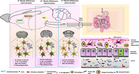
Clinical diagnosis of Parkinson's disease (PD) occurs typically when a substantial proportion of dopaminergic neurons in the substantia nigra (SN) already died, and the first motor symptoms appear. Therefore, tools enabling the early diagnosis of PD are essential to identify early-stage PD patients in which neuroprotective treatments could have a significant impact. Here, we test the utility and sensitivity of the diffusion kurtosis imaging (DKI) in detecting progressive microstructural changes in several brain regions of mice exposed to chronic intragastric administration of rotenone, a mouse model that mimics the spatiotemporal progression of PD-like pathology from the ENS to the SN as described by Braak's staging. Our results show that DKI, especially kurtosis, can detect the progression of pathology-associated changes throughout the CNS. Increases in mean kurtosis were first observed in the dorsal motor nucleus of the vagus (DMV) after 2 months of exposure to rotenone and before the loss of dopaminergic neurons in the SN occurred. Remarkably, we also show that limited exposure to rotenone for 2 months is enough to trigger the progression of the disease in the absence of the environmental toxin, thus suggesting that once the first pathological changes in one region appear, they can self-perpetuate and progress within the CNS. Overall, our results show that DKI can be a useful radiological marker for the early detection and monitoring of PD pathology progression in patients with the potential to improve the clinical diagnosis and the development of neuroprotective treatments.
中文翻译:

扩散峰度成像检测帕金森病口服鱼藤酮小鼠模型病理变化的时间依赖性进展
帕金森病 (PD) 的临床诊断通常发生在黑质 (SN) 中相当大比例的多巴胺能神经元已经死亡,并且出现最初的运动症状时。因此,能够早期诊断 PD 的工具对于识别神经保护治疗可能产生重大影响的早期 PD 患者至关重要。在这里,我们测试了扩散峰度成像 (DKI) 在检测暴露于慢性胃内注射鱼藤酮的小鼠的几个大脑区域的渐进性微观结构变化方面的效用和敏感性,鱼藤酮是一种模拟 PD 样病理学时空进展的小鼠模型。 ENS 到 SN,如 Braak 的分期所述。我们的结果表明,DKI,尤其是峰度,可以检测整个 CNS 病理相关变化的进展。在暴露于鱼藤酮 2 个月后和 SN 中多巴胺能神经元丢失之前,首先在迷走神经背运动核 (DMV) 中观察到平均峰度的增加。值得注意的是,我们还表明,在没有环境毒素的情况下,2 个月的有限暴露于鱼藤酮足以引发疾病的进展,这表明一旦某个区域出现第一个病理变化,它们可以自我延续和发展在中枢神经系统内。总的来说,我们的结果表明,DKI 可以作为一种有用的放射学标志物,用于早期检测和监测患者的 PD 病理进展,有可能改善临床诊断和神经保护治疗的发展。在暴露于鱼藤酮 2 个月后和 SN 中多巴胺能神经元丢失之前,首先在迷走神经背运动核 (DMV) 中观察到平均峰度的增加。值得注意的是,我们还表明,在没有环境毒素的情况下,2 个月的有限暴露于鱼藤酮足以引发疾病的进展,这表明一旦某个区域出现第一个病理变化,它们可以自我延续和发展在中枢神经系统内。总的来说,我们的结果表明,DKI 可以作为一种有用的放射学标志物,用于早期检测和监测患者的 PD 病理进展,有可能改善临床诊断和神经保护治疗的发展。在暴露于鱼藤酮 2 个月后和 SN 中多巴胺能神经元丢失之前,首先在迷走神经背运动核 (DMV) 中观察到平均峰度的增加。值得注意的是,我们还表明,在没有环境毒素的情况下,2 个月的有限暴露于鱼藤酮足以引发疾病的进展,这表明一旦某个区域出现第一个病理变化,它们可以自我延续和发展在中枢神经系统内。总的来说,我们的结果表明,DKI 可以作为一种有用的放射学标志物,用于早期检测和监测患者的 PD 病理进展,有可能改善临床诊断和神经保护治疗的发展。
更新日期:2021-08-12
中文翻译:
扩散峰度成像检测帕金森病口服鱼藤酮小鼠模型病理变化的时间依赖性进展
帕金森病 (PD) 的临床诊断通常发生在黑质 (SN) 中相当大比例的多巴胺能神经元已经死亡,并且出现最初的运动症状时。因此,能够早期诊断 PD 的工具对于识别神经保护治疗可能产生重大影响的早期 PD 患者至关重要。在这里,我们测试了扩散峰度成像 (DKI) 在检测暴露于慢性胃内注射鱼藤酮的小鼠的几个大脑区域的渐进性微观结构变化方面的效用和敏感性,鱼藤酮是一种模拟 PD 样病理学时空进展的小鼠模型。 ENS 到 SN,如 Braak 的分期所述。我们的结果表明,DKI,尤其是峰度,可以检测整个 CNS 病理相关变化的进展。在暴露于鱼藤酮 2 个月后和 SN 中多巴胺能神经元丢失之前,首先在迷走神经背运动核 (DMV) 中观察到平均峰度的增加。值得注意的是,我们还表明,在没有环境毒素的情况下,2 个月的有限暴露于鱼藤酮足以引发疾病的进展,这表明一旦某个区域出现第一个病理变化,它们可以自我延续和发展在中枢神经系统内。总的来说,我们的结果表明,DKI 可以作为一种有用的放射学标志物,用于早期检测和监测患者的 PD 病理进展,有可能改善临床诊断和神经保护治疗的发展。在暴露于鱼藤酮 2 个月后和 SN 中多巴胺能神经元丢失之前,首先在迷走神经背运动核 (DMV) 中观察到平均峰度的增加。值得注意的是,我们还表明,在没有环境毒素的情况下,2 个月的有限暴露于鱼藤酮足以引发疾病的进展,这表明一旦某个区域出现第一个病理变化,它们可以自我延续和发展在中枢神经系统内。总的来说,我们的结果表明,DKI 可以作为一种有用的放射学标志物,用于早期检测和监测患者的 PD 病理进展,有可能改善临床诊断和神经保护治疗的发展。在暴露于鱼藤酮 2 个月后和 SN 中多巴胺能神经元丢失之前,首先在迷走神经背运动核 (DMV) 中观察到平均峰度的增加。值得注意的是,我们还表明,在没有环境毒素的情况下,2 个月的有限暴露于鱼藤酮足以引发疾病的进展,这表明一旦某个区域出现第一个病理变化,它们可以自我延续和发展在中枢神经系统内。总的来说,我们的结果表明,DKI 可以作为一种有用的放射学标志物,用于早期检测和监测患者的 PD 病理进展,有可能改善临床诊断和神经保护治疗的发展。


 京公网安备 11010802027423号