当前位置:
X-MOL 学术
›
J. Neurosci. Res.
›
论文详情
Our official English website, www.x-mol.net, welcomes your
feedback! (Note: you will need to create a separate account there.)
A multielectrode array-based hypoxia model for the analysis of electrical activity in murine retinae
Journal of Neuroscience Research ( IF 2.9 ) Pub Date : 2021-06-10 , DOI: 10.1002/jnr.24899 Claudia Ingensiep 1 , Kim Schaffrath 1 , Bernd Denecke 2 , Peter Walter 1 , Sandra Johnen 1
Journal of Neuroscience Research ( IF 2.9 ) Pub Date : 2021-06-10 , DOI: 10.1002/jnr.24899 Claudia Ingensiep 1 , Kim Schaffrath 1 , Bernd Denecke 2 , Peter Walter 1 , Sandra Johnen 1
Affiliation
|
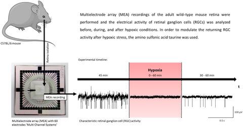
Several eye diseases, for example, retinal artery occlusion, diabetic retinopathy, and glaucoma, are associated with retinal hypoxia. The lack of oxygen in the retina, especially in retinal ganglion cells (RGCs), causes cell damage up to cell degeneration and leads to blindness. Using multielectrode array recordings, an ex vivo hypoxia acute model was established to analyze the electrical activity of murine wild-type retinae under hypoxic stress conditions. Hypoxia was induced by exchanging the perfusion with oxygen-saturated medium by nitrogen-saturated medium. Hypoxic periods of 0 min (control) up to 60 min were tested on the retinae of adult female C57BL/6J mice. The electrical RGC activity vanished during hypoxia, but conditionally returned after the reestablishment of conventional test conditions. With increasing duration of hypoxia, the returning RGC activity decreased. After a hypoxic period of 30 min and a subsequent recovery time of 30 min, 59.43 ± 11.35% of the initially active channels showed a restored RGC activity. The survival rate of retinal cells after hypoxic stress was analyzed by a live/dead staining assay using two-photon laser scanning microscopy. For detailed information about molecular changes caused by hypoxia, a microarray gene expression analysis was performed. Furthermore, the effect of 2-aminoethanesulfonic acid (taurine, 1 mM) on retinae under hypoxic stress was tested. Treatment with taurine resulted in an increase in the RGC response rate after hypoxia and also increased the survival rate of retinal cells under hypoxic stress, confirming its potential as promising candidate for neuroprotective therapies of eye diseases.
中文翻译:

用于分析小鼠视网膜电活动的基于多电极阵列的缺氧模型
一些眼部疾病,例如视网膜动脉阻塞、糖尿病性视网膜病变和青光眼,与视网膜缺氧有关。视网膜中缺氧,尤其是视网膜神经节细胞 (RGC) 中的缺氧,会导致细胞损伤直至细胞退化并导致失明。使用多电极阵列记录,离体建立缺氧急性模型,分析小鼠野生型视网膜在缺氧应激条件下的电活动。通过用氮饱和培养基与氧饱和培养基交换灌注来诱导缺氧。在成年雌性 C57BL/6J 小鼠的视网膜上测试了 0 分钟(对照)至 60 分钟的缺氧期。RGC 电活动在缺氧期间消失,但在重新建立常规测试条件后有条件地恢复。随着缺氧时间的增加,返回的 RGC 活性降低。在 30 分钟的缺氧期和随后的 30 分钟恢复时间后,59.43 ± 11.35% 的初始活动通道显示出恢复的 RGC 活动。使用双光子激光扫描显微镜通过活/死染色测定分析缺氧应激后视网膜细胞的存活率。有关缺氧引起的分子变化的详细信息,进行了微阵列基因表达分析。此外,测试了 2-氨基乙磺酸(牛磺酸,1 mM)在缺氧应激下对视网膜的影响。牛磺酸治疗导致缺氧后 RGC 反应率增加,并且还增加了缺氧应激下视网膜细胞的存活率,证实了其作为眼部疾病神经保护疗法的有希望的候选者的潜力。
更新日期:2021-08-11
中文翻译:
用于分析小鼠视网膜电活动的基于多电极阵列的缺氧模型
一些眼部疾病,例如视网膜动脉阻塞、糖尿病性视网膜病变和青光眼,与视网膜缺氧有关。视网膜中缺氧,尤其是视网膜神经节细胞 (RGC) 中的缺氧,会导致细胞损伤直至细胞退化并导致失明。使用多电极阵列记录,离体建立缺氧急性模型,分析小鼠野生型视网膜在缺氧应激条件下的电活动。通过用氮饱和培养基与氧饱和培养基交换灌注来诱导缺氧。在成年雌性 C57BL/6J 小鼠的视网膜上测试了 0 分钟(对照)至 60 分钟的缺氧期。RGC 电活动在缺氧期间消失,但在重新建立常规测试条件后有条件地恢复。随着缺氧时间的增加,返回的 RGC 活性降低。在 30 分钟的缺氧期和随后的 30 分钟恢复时间后,59.43 ± 11.35% 的初始活动通道显示出恢复的 RGC 活动。使用双光子激光扫描显微镜通过活/死染色测定分析缺氧应激后视网膜细胞的存活率。有关缺氧引起的分子变化的详细信息,进行了微阵列基因表达分析。此外,测试了 2-氨基乙磺酸(牛磺酸,1 mM)在缺氧应激下对视网膜的影响。牛磺酸治疗导致缺氧后 RGC 反应率增加,并且还增加了缺氧应激下视网膜细胞的存活率,证实了其作为眼部疾病神经保护疗法的有希望的候选者的潜力。


 京公网安备 11010802027423号