当前位置:
X-MOL 学术
›
J. Neuroendocrinol.
›
论文详情
Our official English website, www.x-mol.net, welcomes your
feedback! (Note: you will need to create a separate account there.)
Morphological evidence indicates a role for microglia in shaping the PCOS-like brain
Journal of Neuroendocrinology ( IF 3.3 ) Pub Date : 2021-06-04 , DOI: 10.1111/jne.12999 Aisha Sati 1 , Melanie Prescott 1 , Sarah Holland 1 , Christine L Jasoni 1 , Elodie Desroziers 1 , Rebecca E Campbell 1
Journal of Neuroendocrinology ( IF 3.3 ) Pub Date : 2021-06-04 , DOI: 10.1111/jne.12999 Aisha Sati 1 , Melanie Prescott 1 , Sarah Holland 1 , Christine L Jasoni 1 , Elodie Desroziers 1 , Rebecca E Campbell 1
Affiliation
|
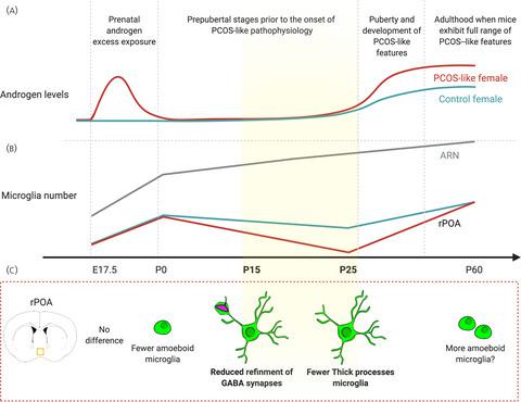
Although polycystic ovary syndrome (PCOS) is the most common cause of anovulatory infertility worldwide, the aetiology of the disorder remains poorly defined. Animal-based evidence highlights the brain as a prime suspect in both the development and maintenance of PCOS. Prenatally androgenised (PNA) models of PCOS exhibit excessive GABAergic wiring associated with PCOS-like reproductive deficits in adulthood, with aberrant brain wiring detected as early as postnatal day (P) 25, prior to disease onset, in the PNA mouse. The mechanisms underlying this aberrant brain wiring remain unknown. Microglia, the immune cells of the brain, are regulators of neuronal wiring across development, mediating both the formation and removal of neuronal inputs. Here, we tested the hypothesis that microglia play a role in the excessive GABAergic wiring that leads to PCOS-like features in the PNA brain. Using specific immunolabelling, microglia number and morphology associated with activation states were analysed in PNA and vehicle-treated controls across developmental timepoints, including embryonic day 17.5, P0, P25 and P60 (n = 7-14 per group), and in two regions of the hypothalamus implicated in fertility regulation. At P0, fewer amoeboid microglia were observed in the rostral preoptic area (rPOA) of PNA mice. However, the greatest changes were observed at P25, with PNA mice exhibiting fewer total microglia, and specifically fewer “sculpting” microglia, in the rPOA. Based on these findings, we assessed microglia-mediated refinement of GABAergic synaptic terminals at two developmental stages of peak synaptic refinement: P7 and P15 (n = 7 per group). PNA mice showed a reduction in the uptake of GABAergic synaptic material at P15. These findings reveal time-specific changes in the microglia population and refinement of GABAergic inputs in a mouse model of PCOS driven by prenatal androgen excess and suggest a role for microglia in shaping the atypical brain wiring associated with the development of PCOS features.
中文翻译:

形态学证据表明小胶质细胞在塑造 PCOS 样大脑中的作用
尽管多囊卵巢综合征 (PCOS) 是全世界无排卵性不孕症的最常见原因,但该疾病的病因仍然不明确。基于动物的证据强调大脑是 PCOS 发展和维持的主要嫌疑人。PCOS 的产前雄激素 (PNA) 模型表现出与成年期 PCOS 样生殖缺陷相关的过度 GABAergic 布线,早在 PNA 小鼠中,早在疾病发作前的出生后第 (P) 25 天 (P) 就检测到异常的脑布线。这种异常大脑接线背后的机制仍然未知。小胶质细胞是大脑的免疫细胞,是整个发育过程中神经元布线的调节剂,介导神经元输入的形成和去除。这里,我们检验了小胶质细胞在导致 PNA 大脑中 PCOS 样特征的过度 GABAergic 布线中发挥作用的假设。使用特异性免疫标记,在 PNA 和载体处理的对照中跨发育时间点分析与激活状态相关的小胶质细胞数量和形态,包括胚胎第 17.5 天、P0、P25 和 P60(每组 n = 7-14),以及两个区域下丘脑参与生育调节。在 P0 时,在 PNA 小鼠的头端视前区 (rPOA) 中观察到较少的变形小胶质细胞。然而,在 P25 观察到最大的变化,PNA 小鼠在 rPOA 中表现出更少的总小胶质细胞,特别是更少的“雕刻”小胶质细胞。基于这些发现,我们在突触细化峰值的两个发育阶段评估了小胶质细胞介导的 GABA 能突触末端细化:P7 和 P15(每组 n = 7)。PNA 小鼠在 P15 显示 GABA 能突触材料的摄取减少。这些发现揭示了产前雄激素过多驱动的 PCOS 小鼠模型中小胶质细胞群的时间特异性变化和 GABA 能输入的细化,并表明小胶质细胞在塑造与 PCOS 特征发展相关的非典型脑布线中的作用。
更新日期:2021-06-04
中文翻译:
形态学证据表明小胶质细胞在塑造 PCOS 样大脑中的作用
尽管多囊卵巢综合征 (PCOS) 是全世界无排卵性不孕症的最常见原因,但该疾病的病因仍然不明确。基于动物的证据强调大脑是 PCOS 发展和维持的主要嫌疑人。PCOS 的产前雄激素 (PNA) 模型表现出与成年期 PCOS 样生殖缺陷相关的过度 GABAergic 布线,早在 PNA 小鼠中,早在疾病发作前的出生后第 (P) 25 天 (P) 就检测到异常的脑布线。这种异常大脑接线背后的机制仍然未知。小胶质细胞是大脑的免疫细胞,是整个发育过程中神经元布线的调节剂,介导神经元输入的形成和去除。这里,我们检验了小胶质细胞在导致 PNA 大脑中 PCOS 样特征的过度 GABAergic 布线中发挥作用的假设。使用特异性免疫标记,在 PNA 和载体处理的对照中跨发育时间点分析与激活状态相关的小胶质细胞数量和形态,包括胚胎第 17.5 天、P0、P25 和 P60(每组 n = 7-14),以及两个区域下丘脑参与生育调节。在 P0 时,在 PNA 小鼠的头端视前区 (rPOA) 中观察到较少的变形小胶质细胞。然而,在 P25 观察到最大的变化,PNA 小鼠在 rPOA 中表现出更少的总小胶质细胞,特别是更少的“雕刻”小胶质细胞。基于这些发现,我们在突触细化峰值的两个发育阶段评估了小胶质细胞介导的 GABA 能突触末端细化:P7 和 P15(每组 n = 7)。PNA 小鼠在 P15 显示 GABA 能突触材料的摄取减少。这些发现揭示了产前雄激素过多驱动的 PCOS 小鼠模型中小胶质细胞群的时间特异性变化和 GABA 能输入的细化,并表明小胶质细胞在塑造与 PCOS 特征发展相关的非典型脑布线中的作用。


 京公网安备 11010802027423号