Journal of Physics and Chemistry of Solids ( IF 4.3 ) Pub Date : 2021-06-05 , DOI: 10.1016/j.jpcs.2021.110207 Ababay Ketema Worku , Delele Worku Ayele , Nigus Gabbiye Habtu , Minbale Admas Teshager , Zerihun Getahun Workineh
|
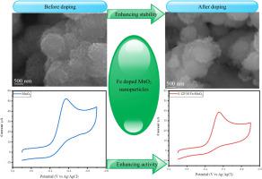
In this study, 3D flower-like MnO2 and Fe–MnO2 nanoparticles were developed using a co-perception method. In the doping manner, the concentration of Fe was raised from 0.025 M to 0.125 M in levels of 0.025 M. The prepared electrocatalysts were calcined at 500 °C. Different analytical techniques, particularly XRD, SEM, UV–Vis, FTIR, TGA/DTA, CV, ICP-OES, BET, and LSV were utilized to analyze the structure, morphology, optical, thermal, and electrochemical activity of 3D flower-like MnO2 and Fe–MnO2 nanoparticles. XRD analysis showed the phase change from ε to λ and the phase of MnO2 and the crystallite sizes of un-doped MnO2 and Fe–MnO2 were between 1.18 and 2.25 nm. Morphological examinations indicated that pure MnO2 and Fe-doped MnO2 have spherical-like flake-flower and agglomerated nanoparticle architectures, respectively. The BET surface area of 0.125 M Fe–MnO2 nanoparticles is 288.2 m2g-1. The bandgap energies of MnO2, 0.05 M Fe-doped MnO2, and 0.125 M Fe-doped MnO2 nanoparticles were determined to be 0.57 eV, 0.18 eV, and 0.14 eV, respectively as evident from UV–Vis. The presence of M − O bonds (M = Mn, Fe) was analyzed by FTIR spectroscopy. The doped MnO2 electrocatalyst shows improved thermal properties owing to the doping effect of iron. The results of CV and LSV measurements reveal that the Fe–MnO2 nanoparticles have an excellent catalytic performance toward the ORR under 0.1 M KOH alkaline conditions.
中文翻译:
通过铁掺杂增强ε-MnO 2纳米颗粒的氧还原反应活性
在这项研究中,使用共同感知方法开发了3D 花状 MnO 2和 Fe-MnO 2纳米粒子。在掺杂方式中,Fe 的浓度从 0.025 M 提高到 0.125 M,水平为 0.025 M。制备的电催化剂在 500 ° C下煅烧 。不同的分析技术,特别是 XRD、SEM、UV-Vis、FTIR、TGA/ DTA、CV、ICP-OES、BET 和 LSV 被用来分析 3D 花状 MnO 2和 Fe-MnO 2纳米粒子的结构、形态、光学、热和电化学活性。XRD分析表明从ε到λ的相变以及MnO 2的相和未掺杂的MnO 2的微晶尺寸和 Fe-MnO 2介于 1.18 和 2.25 nm 之间。形态学检查表明纯MnO 2和Fe掺杂的MnO 2 分别具有球形片状花和团聚纳米颗粒结构。0.125 M Fe-MnO 2纳米颗粒的BET表面积为288.2 m 2 g -1。MnO 2、0.05 M Fe 掺杂的MnO 2和0.125 M Fe 掺杂的MnO 2纳米粒子的带隙能量分别测定为0.57 eV、0.18 eV 和0.14 eV,从UV-Vis 可见。M - O 键 (M = Mn, Fe) 的存在通过 FTIR 光谱分析。掺杂的MnO 2由于铁的掺杂作用,电催化剂显示出改善的热性能。CV 和 LSV 测量结果表明,Fe-MnO 2纳米颗粒在 0.1 M KOH 碱性条件下对 ORR 具有优异的催化性能。


 京公网安备 11010802027423号