当前位置:
X-MOL 学术
›
Immunology
›
论文详情
Our official English website, www.x-mol.net, welcomes your feedback! (Note: you will need to create a separate account there.)
Neutralizing interferon-α blocks inflammation-mediated vascular injury via PI3K and AMPK in systemic lupus erythematosus
Immunology ( IF 6.4 ) Pub Date : 2021-06-02 , DOI: 10.1111/imm.13379 Xuewei Ding 1, 2 , Wei Xiang 3 , Ren Yi 1, 4 , Xiaoyan Huang 3 , Qiuyu Lin 3 , Xiaojie He 1, 2
Immunology ( IF 6.4 ) Pub Date : 2021-06-02 , DOI: 10.1111/imm.13379 Xuewei Ding 1, 2 , Wei Xiang 3 , Ren Yi 1, 4 , Xiaoyan Huang 3 , Qiuyu Lin 3 , Xiaojie He 1, 2
Affiliation
|
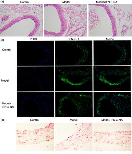
Plasmacytoid dendritic cells (pDCs) play a key role in the initiation and amplification of systemic lupus erythematosus (SLE)-associated vascular injury. In this study, we found that dsDNA induced dose- and time-dependent increase in IFN-α and Toll-like receptor 7 (TLR7), TLR9 and IRF7 expression in pDCs. Co-cultured circulating endothelial cells (ECs) with activated pDCs significantly decreased proliferation, tube formation and migration in ECs. The elevated level of cellular IFN-α increased cell adhesion, promoted cell apoptosis, induced cell senescence and arrested cells at G0/G1 phase of endothelial progenitor cells (EPCs). Additionally, the co-culture system activated MAPK and inactivated PI3K. Pristane was used to establish a in vivo SLE-like mouse model. Importantly, we showed that INF-α-neutralizing antibody (IFN-α-NA) rescued all the changes induced by IFN-α in vitro and prevented vascular injury in pristane-induced SLE model in vivo. In conclusion, we confirmed that activated pDCs promoted vascular damage and the dysfunction of ECs/EPCs via IFN-α production. IFN-α-neutralizing antibody may be a clinical implication for preventing vascular injury. PI3K signalling and AMPK signalling were associated with SLE-associated vascular functions.
中文翻译:

在系统性红斑狼疮中中和干扰素-α 通过 PI3K 和 AMPK 阻断炎症介导的血管损伤
浆细胞样树突状细胞 (pDC) 在系统性红斑狼疮 (SLE) 相关血管损伤的启动和放大中起关键作用。在这项研究中,我们发现 dsDNA 诱导 pDC 中 IFN-α 和 Toll 样受体 7 (TLR7)、TLR9 和 IRF7 表达的剂量和时间依赖性增加。与活化的 pDCs 共培养的循环内皮细胞 (ECs) 显着降低了 ECs 中的增殖、管形成和迁移。细胞内 IFN-α 水平的升高增加了细胞粘附,促进细胞凋亡,诱导细胞衰老并在内皮祖细胞 (EPCs) 的 G0/G1 期停滞细胞。此外,共培养系统激活了 MAPK 并灭活了 PI3K。使用 Pristane 建立体内SLE 样鼠标模型。重要的是,我们发现 INF-α 中和抗体 (IFN-α-NA)在体外挽救了由 IFN-α 诱导的所有变化,并防止了体内 pristane 诱导的 SLE 模型中的血管损伤。总之,我们证实活化的 pDCs 通过产生 IFN-α 促进血管损伤和 ECs/EPCs 的功能障碍。IFN-α中和抗体可能是预防血管损伤的临床意义。PI3K 信号和 AMPK 信号与 SLE 相关的血管功能相关。
更新日期:2021-06-02
中文翻译:
在系统性红斑狼疮中中和干扰素-α 通过 PI3K 和 AMPK 阻断炎症介导的血管损伤
浆细胞样树突状细胞 (pDC) 在系统性红斑狼疮 (SLE) 相关血管损伤的启动和放大中起关键作用。在这项研究中,我们发现 dsDNA 诱导 pDC 中 IFN-α 和 Toll 样受体 7 (TLR7)、TLR9 和 IRF7 表达的剂量和时间依赖性增加。与活化的 pDCs 共培养的循环内皮细胞 (ECs) 显着降低了 ECs 中的增殖、管形成和迁移。细胞内 IFN-α 水平的升高增加了细胞粘附,促进细胞凋亡,诱导细胞衰老并在内皮祖细胞 (EPCs) 的 G0/G1 期停滞细胞。此外,共培养系统激活了 MAPK 并灭活了 PI3K。使用 Pristane 建立体内SLE 样鼠标模型。重要的是,我们发现 INF-α 中和抗体 (IFN-α-NA)在体外挽救了由 IFN-α 诱导的所有变化,并防止了体内 pristane 诱导的 SLE 模型中的血管损伤。总之,我们证实活化的 pDCs 通过产生 IFN-α 促进血管损伤和 ECs/EPCs 的功能障碍。IFN-α中和抗体可能是预防血管损伤的临床意义。PI3K 信号和 AMPK 信号与 SLE 相关的血管功能相关。


 京公网安备 11010802027423号