当前位置:
X-MOL 学术
›
Microsc. Res. Tech.
›
论文详情
Our official English website, www.x-mol.net, welcomes your
feedback! (Note: you will need to create a separate account there.)
Analysis of enamel/restoration interface submitted cariogenic challenge and fluoride release
Microscopy Research and Technique ( IF 2.0 ) Pub Date : 2021-05-28 , DOI: 10.1002/jemt.23844 Raquel Viana Rodrigues 1, 2 , Camila Sobral Sampaio 1, 3 , Aline Carvalho Girotto 1 , Caroline Paiuta Pinhatti 4 , Alexsandra Shizue Iwamoto 4 , Anderson Zanardi de Freitas 5 , Gláucia Maria Bovi Ambrosano 4 , Regina Maria Puppin-Rontani 4 , Fernanda Miori Pascon 4
Microscopy Research and Technique ( IF 2.0 ) Pub Date : 2021-05-28 , DOI: 10.1002/jemt.23844 Raquel Viana Rodrigues 1, 2 , Camila Sobral Sampaio 1, 3 , Aline Carvalho Girotto 1 , Caroline Paiuta Pinhatti 4 , Alexsandra Shizue Iwamoto 4 , Anderson Zanardi de Freitas 5 , Gláucia Maria Bovi Ambrosano 4 , Regina Maria Puppin-Rontani 4 , Fernanda Miori Pascon 4
Affiliation
|
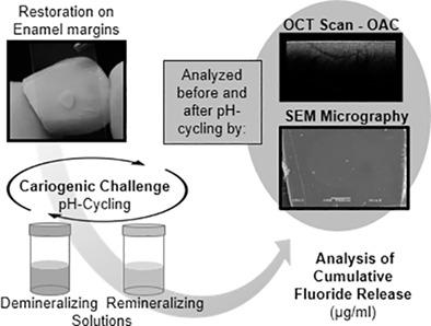
The treatment of high-risk patients still is a challenge. The understanding and development non-invasive, non-destructive, and non-ionizing techniques, can help to guide the treatment and the diagnosis of primary and recurrent caries. The present study evaluated the behavior of enamel/restoration interface after a cariogenic challenge by Fourier domain optical coherence tomography (FD-OCT), scanning electron microscopy (SEM) examination, and the fluoride release of the different restorative materials. Cavities (1.5 × 0.5 mm) were performed in enamel surface and divided into groups (n = 8): glass ionomer cement (GIC), resin-modified glass ionomer cement (RMGIC), and resin composite (RC). The samples were submitted to pH-cycling, and the solutions analyzed for cumulative fluoride by ion-analyzer. The morphology was analyzed by SEM through replicas. The optical attenuation coefficient (OAC) was calculated through exponential decay from the images generated by FD-OCT. Data were analyzed considering α = 0.05. OAC values increased for all groups after pH-cycling indicating demineralization (p < .05). Considering the remineralizing solution, RMGIC presented higher fluoride release rate, followed by GIC, while RC did not release any fluoride. Yet for the demineralizing solution, RMGIC and GIC released similar fluoride rates, overcoming RC (p < .05). Micrographs revealed no changes on the restorations margins, although enamel detachment was observed for RC and GIC after pH-cycling.
中文翻译:

牙釉质/修复界面提交的致龋挑战和氟化物释放的分析
高危患者的治疗仍然是一个挑战。了解和发展非侵入性、非破坏性、非电离技术,有助于指导原发性和复发性龋病的治疗和诊断。本研究通过傅里叶域光学相干断层扫描 (FD-OCT)、扫描电子显微镜 (SEM) 检测和不同修复材料的氟化物释放评估了牙釉质/修复界面在致龋挑战后的行为。在牙釉质表面进行腔(1.5 × 0.5 mm)并分成组(n = 8):玻璃离子水门汀 (GIC)、树脂改性玻璃离子水门汀 (RMGIC) 和树脂复合材料 (RC)。将样品进行 pH 循环,并通过离子分析仪分析溶液的累积氟化物。通过复制品通过SEM分析形态。光学衰减系数 (OAC) 是通过 FD-OCT 生成的图像的指数衰减来计算的。考虑α = 0.05分析数据。pH 循环后所有组的 OAC 值均增加,表明脱矿质 ( p < .05)。考虑到再矿化溶液,RMGIC 呈现出较高的氟化物释放率,其次是 GIC,而 RC 没有释放任何氟化物。然而,对于脱矿溶液,RMGIC 和 GIC 释放了相似的氟化物率,克服了 RC ( p < .05)。显微照片显示修复体边缘没有变化,尽管在 pH 循环后观察到 RC 和 GIC 的牙釉质脱离。
更新日期:2021-05-28
中文翻译:
牙釉质/修复界面提交的致龋挑战和氟化物释放的分析
高危患者的治疗仍然是一个挑战。了解和发展非侵入性、非破坏性、非电离技术,有助于指导原发性和复发性龋病的治疗和诊断。本研究通过傅里叶域光学相干断层扫描 (FD-OCT)、扫描电子显微镜 (SEM) 检测和不同修复材料的氟化物释放评估了牙釉质/修复界面在致龋挑战后的行为。在牙釉质表面进行腔(1.5 × 0.5 mm)并分成组(n = 8):玻璃离子水门汀 (GIC)、树脂改性玻璃离子水门汀 (RMGIC) 和树脂复合材料 (RC)。将样品进行 pH 循环,并通过离子分析仪分析溶液的累积氟化物。通过复制品通过SEM分析形态。光学衰减系数 (OAC) 是通过 FD-OCT 生成的图像的指数衰减来计算的。考虑α = 0.05分析数据。pH 循环后所有组的 OAC 值均增加,表明脱矿质 ( p < .05)。考虑到再矿化溶液,RMGIC 呈现出较高的氟化物释放率,其次是 GIC,而 RC 没有释放任何氟化物。然而,对于脱矿溶液,RMGIC 和 GIC 释放了相似的氟化物率,克服了 RC ( p < .05)。显微照片显示修复体边缘没有变化,尽管在 pH 循环后观察到 RC 和 GIC 的牙釉质脱离。


 京公网安备 11010802027423号