当前位置:
X-MOL 学术
›
Brain Pathol.
›
论文详情
Our official English website, www.x-mol.net, welcomes your
feedback! (Note: you will need to create a separate account there.)
MRI and muscle imaging for idiopathic inflammatory myopathies
Brain Pathology ( IF 5.8 ) Pub Date : 2021-05-27 , DOI: 10.1111/bpa.12954 Samuel Malartre 1, 2 , Damien Bachasson 3 , Guillaume Mercy 4 , Elissone Sarkis 1, 2 , Céline Anquetil 1, 2 , Olivier Benveniste 1, 2 , Yves Allenbach 1, 2
Brain Pathology ( IF 5.8 ) Pub Date : 2021-05-27 , DOI: 10.1111/bpa.12954 Samuel Malartre 1, 2 , Damien Bachasson 3 , Guillaume Mercy 4 , Elissone Sarkis 1, 2 , Céline Anquetil 1, 2 , Olivier Benveniste 1, 2 , Yves Allenbach 1, 2
Affiliation
|
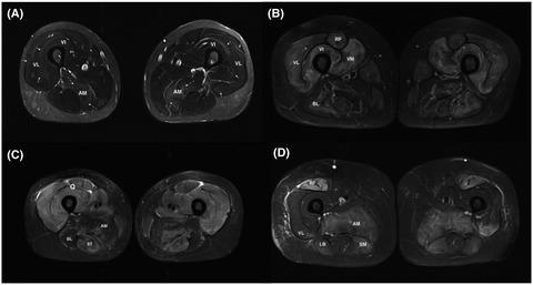
Although idiopathic inflammatory myopathies (IIM) are a heterogeneous group of diseases nearly all patients display muscle inflammation. Originally, muscle biopsy was considered as the gold standard for IIM diagnosis. The development of muscle imaging led to revisiting not only the IIM diagnosis strategy but also the patients’ follow-up. Different techniques have been tested or are in development for IIM including positron emission tomography, ultrasound imaging, ultrasound shear wave elastography, though magnetic resonance imaging (MRI) remains the most widely used technique in routine. Whereas guidelines on muscle imaging in myositis are lacking here we reviewed the relevance of muscle imaging for both diagnosis and myositis patients’ follow-up. We propose recommendations about when and how to perform MRI on myositis patients, and we describe new techniques that are under development.
中文翻译:

特发性炎症性肌病的 MRI 和肌肉成像
尽管特发性炎症性肌病 (IIM) 是一组异质性疾病,但几乎所有患者都表现出肌肉炎症。最初,肌肉活检被认为是 IIM 诊断的金标准。肌肉影像学的发展不仅导致重新审视 IIM 诊断策略,还重新审视患者的随访。IIM 已经测试或正在开发不同的技术,包括正电子发射断层扫描、超声成像、超声剪切波弹性成像,尽管磁共振成像 (MRI) 仍然是常规中使用最广泛的技术。虽然这里缺乏关于肌炎肌肉成像的指南,但我们回顾了肌肉成像对诊断和肌炎患者随访的相关性。我们提出了关于何时以及如何对肌炎患者进行 MRI 的建议,
更新日期:2021-05-27
中文翻译:
特发性炎症性肌病的 MRI 和肌肉成像
尽管特发性炎症性肌病 (IIM) 是一组异质性疾病,但几乎所有患者都表现出肌肉炎症。最初,肌肉活检被认为是 IIM 诊断的金标准。肌肉影像学的发展不仅导致重新审视 IIM 诊断策略,还重新审视患者的随访。IIM 已经测试或正在开发不同的技术,包括正电子发射断层扫描、超声成像、超声剪切波弹性成像,尽管磁共振成像 (MRI) 仍然是常规中使用最广泛的技术。虽然这里缺乏关于肌炎肌肉成像的指南,但我们回顾了肌肉成像对诊断和肌炎患者随访的相关性。我们提出了关于何时以及如何对肌炎患者进行 MRI 的建议,


 京公网安备 11010802027423号