当前位置:
X-MOL 学术
›
ACS Photonics
›
论文详情
Our official English website, www.x-mol.net, welcomes your
feedback! (Note: you will need to create a separate account there.)
Cost-Effective Live Cell Structured Illumination Microscopy with Video-Rate Imaging
ACS Photonics ( IF 6.5 ) Pub Date : 2021-05-26 , DOI: 10.1021/acsphotonics.0c01937 Alice Sandmeyer 1 , Mario Lachetta 1 , Hauke Sandmeyer 2 , Wolfgang Hübner 1 , Thomas Huser 1 , Marcel Müller 1, 3
ACS Photonics ( IF 6.5 ) Pub Date : 2021-05-26 , DOI: 10.1021/acsphotonics.0c01937 Alice Sandmeyer 1 , Mario Lachetta 1 , Hauke Sandmeyer 2 , Wolfgang Hübner 1 , Thomas Huser 1 , Marcel Müller 1, 3
Affiliation
|
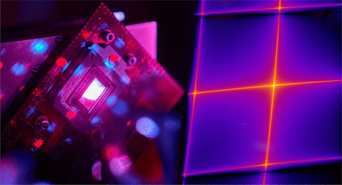
Optical nanoscopy is rapidly gaining momentum in the life sciences. Current instruments are, however, often large and expensive, and there is a substantial delay between raw data collection and super-resolved image display. Here, we describe the implementation of a compact, cost-effective, high-speed, structured illumination microscope (SIM), which allows for video-rate super-resolved image reconstructions at imaging rates up to 60 Hz. The instrument is based on a digital micromirror device (DMD) and a global-shutter camera, which enables faster pattern cycles and higher duty cycles than commonly used liquid crystal-based spatial light modulators. In order to utilize a DMD for creating illumination patterns by the coherent superposition of laser beams, we carefully studied its blazed grating effect Through both simulation and experimental determination of system parameters, we identified and optimized its alignment for optimal SIM pattern contrast. Raw image data are collected using inexpensive industry-grade CMOS cameras, while a parallel-computing platform allowed us to reconstruct and visualize living cells in real time. We demonstrate the performance of this system by imaging submicron-sized fluorescent beads diffusing in an aqueous solution, resolving bead–bead interactions in real time. We show that the system is sensitive enough to image intracellular vesicles labeled with fluorescent proteins in fixed cells. We also image dynamic fluctuations of the endoplasmic reticulum (ER), as well as the movement of mitochondria in living osteosarcoma cells, where the cellular organelles are labeled with live cell fluorescent stains.
中文翻译:

具有视频速率成像的经济高效的活细胞结构照明显微镜
光学纳米技术在生命科学领域的发展势头迅猛。然而,当前的仪器通常庞大且昂贵,并且在原始数据收集和超分辨率图像显示之间存在大量延迟。在这里,我们描述了一种紧凑、经济、高速、结构化照明显微镜 (SIM) 的实现,它允许以高达 60 Hz 的成像速率进行视频速率超分辨率图像重建。该仪器基于数字微镜器件 (DMD) 和全局快门相机,与常用的基于液晶的空间光调制器相比,可实现更快的图案周期和更高的占空比。为了利用 DMD 通过激光束的相干叠加来创建照明图案,我们仔细研究了它的闪耀光栅效应通过系统参数的模拟和实验确定,我们确定并优化了它的对齐方式,以获得最佳的 SIM 图案对比度。原始图像数据是使用廉价的工业级 CMOS 相机收集的,而并行计算平台使我们能够实时重建和可视化活细胞。我们通过对在水溶液中扩散的亚微米大小的荧光珠进行成像,实时解决珠-珠相互作用来证明该系统的性能。我们表明该系统足够敏感,可以对固定细胞中用荧光蛋白标记的细胞内囊泡进行成像。我们还对内质网 (ER) 的动态波动以及活骨肉瘤细胞中线粒体的运动进行了成像,
更新日期:2021-06-17
中文翻译:
具有视频速率成像的经济高效的活细胞结构照明显微镜
光学纳米技术在生命科学领域的发展势头迅猛。然而,当前的仪器通常庞大且昂贵,并且在原始数据收集和超分辨率图像显示之间存在大量延迟。在这里,我们描述了一种紧凑、经济、高速、结构化照明显微镜 (SIM) 的实现,它允许以高达 60 Hz 的成像速率进行视频速率超分辨率图像重建。该仪器基于数字微镜器件 (DMD) 和全局快门相机,与常用的基于液晶的空间光调制器相比,可实现更快的图案周期和更高的占空比。为了利用 DMD 通过激光束的相干叠加来创建照明图案,我们仔细研究了它的闪耀光栅效应通过系统参数的模拟和实验确定,我们确定并优化了它的对齐方式,以获得最佳的 SIM 图案对比度。原始图像数据是使用廉价的工业级 CMOS 相机收集的,而并行计算平台使我们能够实时重建和可视化活细胞。我们通过对在水溶液中扩散的亚微米大小的荧光珠进行成像,实时解决珠-珠相互作用来证明该系统的性能。我们表明该系统足够敏感,可以对固定细胞中用荧光蛋白标记的细胞内囊泡进行成像。我们还对内质网 (ER) 的动态波动以及活骨肉瘤细胞中线粒体的运动进行了成像,


 京公网安备 11010802027423号