Clinical Spectroscopy Pub Date : 2021-05-26 , DOI: 10.1016/j.clispe.2021.100011 Valentina Notarstefano , Simona Sabbatini , Maurizio Sabbatini , Aldo Arrais , Alessia Belloni , Chiara Pro , Lisa Vaccari , Domenico Osella , Elisabetta Giorgini
|
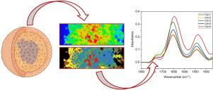
Multicellular spheroids are the new frontier for studying how the tumour micro-environment interferes with drug uptake and response, since they can reproduce a three-dimensional cellular organisation mimicking the behaviour of in vivo solid tissues. In this study, we exploited Focal Plane Array - Fourier Transform Infrared Imaging spectroscopy to characterize the biochemical features, in terms of distribution and composition of the meaningful macromolecules (lipids, proteins, sugars and nucleic acids), of malignant pleural mesothelioma spheroid sections, and, as further extent, to investigate the penetrating effects of cisplatin within the spheroid mass. The hyperspectral imaging analysis evidenced, in untreated spheroids, the occurrence of a replicative outer region and a hypoxic inner one, as suggested by the band area ratios related to lipid alkyl chains (2925/2960) and glycogen (1020/1650), which showed the highest values in the inner region. Moreover, the HCA spectroscopic images showed, after cisplatin treatment, an increase of the band area ratio related to lipid carbonyl ester moiety (1740/2925), suggesting the occurrence of lipid peroxidation; furthermore, the band area ratio related to nucleic acids (1240/1220) revealed a DNA fragmentation along all regions of spheroids that may be related to apoptotic mechanisms, whereas a reduction of the band area ratios related to glycogen and carbohydrates (1020/1650 and 1054/1650, respectively) appeared consistent with an inhibition of cell division. The few spectral differences between the outer and the inner regions of cisplatin-treated spheroids pointed out the diffuse penetrating effect of the drug.
中文翻译:
MSTO-211H 细胞球体模型的高光谱表征:FPA-FTIR 成像方法
多细胞球体是研究肿瘤微环境如何干扰药物摄取和反应的新前沿,因为它们可以重现模仿体内行为的三维细胞组织实体组织。在这项研究中,我们利用焦平面阵列 - 傅里叶变换红外成像光谱来表征恶性胸膜间皮瘤球体切片的生化特征,包括有意义的大分子(脂质、蛋白质、糖和核酸)的分布和组成,以及进一步研究顺铂在球状体中的穿透作用。高光谱成像分析证明,在未经处理的球体中,出现了复制的外部区域和缺氧的内部区域,正如与脂烷基链 (2925/2960) 和糖原 (1020/1650) 相关的带面积比所表明的那样,这表明内部区域的最高值。此外,HCA 光谱图像显示,顺铂处理后,与脂质羰基酯部分相关的带面积比增加(1740/2925),表明脂质过氧化的发生;此外,与核酸相关的带面积比 (1240/1220) 揭示了可能与凋亡机制相关的球体所有区域的 DNA 断裂,而与糖原和碳水化合物相关的带面积比降低 (1020/1650 和1054/1650)似乎与细胞分裂的抑制一致。顺铂处理的球体的外部和内部区域之间的少量光谱差异表明了药物的弥散渗透作用。与核酸相关的条带面积比 (1240/1220) 揭示了可能与细胞凋亡机制相关的球体所有区域的 DNA 断裂,而与糖原和碳水化合物相关的条带面积比 (1020/1650 和 1054/ 1650)似乎与细胞分裂的抑制一致。顺铂处理的球体的外部和内部区域之间的少量光谱差异表明了药物的弥散渗透作用。与核酸相关的条带面积比 (1240/1220) 揭示了可能与细胞凋亡机制相关的球体所有区域的 DNA 断裂,而与糖原和碳水化合物相关的条带面积比 (1020/1650 和 1054/ 1650)似乎与细胞分裂的抑制一致。顺铂处理的球体的外部和内部区域之间的少量光谱差异表明了药物的弥散渗透作用。


 京公网安备 11010802027423号