Computers & Graphics ( IF 2.5 ) Pub Date : 2021-05-24 , DOI: 10.1016/j.cag.2021.05.006 Yong Wan 1 , Holly A Holman 1 , Charles Hansen 1
|
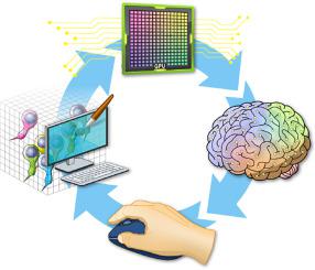
The main objective for understanding fluorescence microscopy data is to investigate and evaluate the fluorescent signal intensity distributions as well as their spatial relationships across multiple channels. The quantitative analysis of 3D fluorescence microscopy data needs interactive tools for researchers to select and focus on relevant biological structures. We developed an interactive tool based on volume visualization techniques and GPU computing for streamlining rapid data analysis. Our main contribution is the implementation of common data quantification functions on streamed volumes, providing interactive analyses on large data without lengthy preprocessing. Data segmentation and quantification are coupled with brushing and executed at an interactive speed. A large volume is partitioned into data bricks, and only user-selected structures are analyzed to constrain the computational load. We designed a framework to assemble a sequence of GPU programs to handle brick borders and stitch analysis results. Our tool was developed in collaboration with domain experts and has been used to identify cell types. We demonstrate a workflow to analyze cells in vestibular epithelia of transgenic mice.
中文翻译:
以细胞精度对来自荧光显微镜的大量数据进行交互式分析
了解荧光显微镜数据的主要目的是研究和评估荧光信号强度分布及其跨多个通道的空间关系。3D 荧光显微镜数据的定量分析需要交互式工具,供研究人员选择和关注相关的生物结构。我们开发了一种基于体可视化技术和 GPU 计算的交互式工具,用于简化快速数据分析。我们的主要贡献是在流卷上实现通用数据量化功能,无需冗长的预处理即可对大数据进行交互式分析。数据分割和量化与刷机相结合,并以交互速度执行。大卷被划分为数据块,并且仅分析用户选择的结构以限制计算负载。我们设计了一个框架来组装一系列 GPU 程序来处理砖边界和缝合分析结果。我们的工具是与领域专家合作开发的,已用于识别细胞类型。我们展示了一个分析转基因小鼠前庭上皮细胞的工作流程。


 京公网安备 11010802027423号