当前位置:
X-MOL 学术
›
NMR Biomed.
›
论文详情
Our official English website, www.x-mol.net, welcomes your
feedback! (Note: you will need to create a separate account there.)
High-contrast osteochondral junction imaging using a 3D dual adiabatic inversion recovery-prepared ultrashort echo time cones sequence
NMR in Biomedicine ( IF 2.7 ) Pub Date : 2021-05-22 , DOI: 10.1002/nbm.4559 Alecio F Lombardi 1, 2 , Hyungseok Jang 1 , Zhao Wei 1 , Saeed Jerban 1 , Mark Wallace 3 , Koichi Masuda 4 , Ya-Jun Ma 1
NMR in Biomedicine ( IF 2.7 ) Pub Date : 2021-05-22 , DOI: 10.1002/nbm.4559 Alecio F Lombardi 1, 2 , Hyungseok Jang 1 , Zhao Wei 1 , Saeed Jerban 1 , Mark Wallace 3 , Koichi Masuda 4 , Ya-Jun Ma 1
Affiliation
|
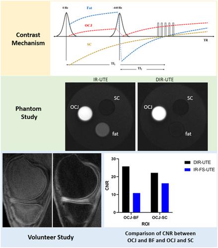
While conventional MRI sequences cannot visualize tissues from the osteochondral junction (OCJ) due to these tissues' short transverse T2/T2* relaxations, ultrashort echo time (UTE) sequences can overcome this limitation. A 2D UTE sequence with a dual adiabatic inversion recovery preparation (DIR-UTE) for selective imaging of short T2 tissues with high contrast has previously been developed, but high sensitivity to eddy currents and aliased out-of-slice excitation make it difficult to image the thin layer of the OCJ in vivo. Here, we combine the DIR scheme with a 3D UTE cones sequence for volumetric imaging of OCJ tissues in vivo, aiming to generate higher OCJ contrast compared with a recently developed single IR-prepared UTE sequence with a fat saturation module (IR-FS-UTE). All sequences were implemented on a 3-T clinical scanner. The DIR-UTE cones sequence combined a 3D UTE cones sequence with two narrow-band adiabatic IR preparation pulses centered on water and fat spectrum frequencies, respectively. The 3D DIR-UTE cones sequence was first applied to a phantom, then to the knees of four healthy volunteers and four patients diagnosed with osteoarthritis and compared with the IR-FS-UTE sequence. In both phantom and volunteer studies, the proposed DIR-UTE cones sequence showed much higher contrast for OCJ imaging than the IR-FS-UTE sequence did. The 3D DIR-UTE cones sequence showed a significantly higher contrast-to-noise ratio between the OCJ and subchondral bone fat (mean, standard deviation [SD]: 25.7 ± 2.3) and between the OCJ and superficial layers of cartilage (mean, SD: 22.2 ± 3.5) compared with the IR-FS-UTE sequence (mean, SD: 10.8 ± 2.5 and 16.3 ± 2.6, respectively). The 3D DIR-UTE cones sequence is feasible for imaging of the OCJ region of the knee in vivo and produces both high resolution and high contrast.
中文翻译:

使用 3D 双绝热反转恢复制备的超短回波时间锥序列的高对比度骨软骨结成像
虽然传统的 MRI 序列由于这些组织的短横向 T 2 /T 2 * 弛豫而无法显示来自骨软骨连接 (OCJ) 的组织,但超短回波时间 (UTE) 序列可以克服这一限制。具有双绝热反转恢复准备 (DIR-UTE) 的 2D UTE 序列,用于短 T 2的选择性成像以前已经开发了具有高对比度的组织,但是对涡流和混叠的层外激发的高灵敏度使得难以在体内对 OCJ 的薄层进行成像。在这里,我们将 DIR 方案与 3D UTE 锥序列相结合,用于体内 OCJ 组织的体积成像,旨在与最近开发的具有脂肪饱和模块的单个 IR 制备的 UTE 序列(IR-FS-UTE)相比产生更高的 OCJ 对比度。 )。所有序列均在 3-T 临床扫描仪上实施。DIR-UTE 锥体序列将 3D UTE 锥体序列与两个分别以水和脂肪光谱频率为中心的窄带绝热 IR 准备脉冲相结合。3D DIR-UTE 锥体序列首先应用于体模,然后到四名健康志愿者和四名被诊断患有骨关节炎的患者的膝盖上,并与 IR-FS-UTE 序列进行比较。在模型和志愿者研究中,建议的 DIR-UTE 锥序列显示 OCJ 成像的对比度比 IR-FS-UTE 序列高得多。3D DIR-UTE 锥体序列显示 OCJ 和软骨下骨脂肪(平均值,标准差 [SD]:25.7 ± 2.3)之间以及 OCJ 和软骨表层之间(平均值,SD : 22.2 ± 3.5) 与 IR-FS-UTE 序列相比(平均值,SD:分别为 10.8 ± 2.5 和 16.3 ± 2.6)。3D DIR-UTE 锥序列可用于体内膝关节 OCJ 区域的成像,并产生高分辨率和高对比度。建议的 DIR-UTE 锥序列显示 OCJ 成像的对比度比 IR-FS-UTE 序列高得多。3D DIR-UTE 锥体序列显示 OCJ 和软骨下骨脂肪(平均值,标准差 [SD]:25.7 ± 2.3)之间以及 OCJ 和软骨表层之间(平均值,SD : 22.2 ± 3.5) 与 IR-FS-UTE 序列相比(平均值,SD:分别为 10.8 ± 2.5 和 16.3 ± 2.6)。3D DIR-UTE 锥序列可用于体内膝关节 OCJ 区域的成像,并产生高分辨率和高对比度。建议的 DIR-UTE 锥序列显示 OCJ 成像的对比度比 IR-FS-UTE 序列高得多。3D DIR-UTE 锥体序列显示 OCJ 和软骨下骨脂肪(平均值,标准差 [SD]:25.7 ± 2.3)之间以及 OCJ 和软骨表层之间(平均值,SD : 22.2 ± 3.5) 与 IR-FS-UTE 序列相比(平均值,SD:分别为 10.8 ± 2.5 和 16.3 ± 2.6)。3D DIR-UTE 锥序列可用于体内膝关节 OCJ 区域的成像,并产生高分辨率和高对比度。3) 以及 OCJ 和软骨表层之间(平均值,SD:22.2 ± 3.5)与 IR-FS-UTE 序列(平均值,SD:分别为 10.8 ± 2.5 和 16.3 ± 2.6)相比。3D DIR-UTE 锥序列可用于体内膝关节 OCJ 区域的成像,并产生高分辨率和高对比度。3) 以及 OCJ 和软骨表层之间(平均值,SD:22.2 ± 3.5)与 IR-FS-UTE 序列(平均值,SD:分别为 10.8 ± 2.5 和 16.3 ± 2.6)相比。3D DIR-UTE 锥序列可用于体内膝关节 OCJ 区域的成像,并产生高分辨率和高对比度。
更新日期:2021-07-02
中文翻译:
使用 3D 双绝热反转恢复制备的超短回波时间锥序列的高对比度骨软骨结成像
虽然传统的 MRI 序列由于这些组织的短横向 T 2 /T 2 * 弛豫而无法显示来自骨软骨连接 (OCJ) 的组织,但超短回波时间 (UTE) 序列可以克服这一限制。具有双绝热反转恢复准备 (DIR-UTE) 的 2D UTE 序列,用于短 T 2的选择性成像以前已经开发了具有高对比度的组织,但是对涡流和混叠的层外激发的高灵敏度使得难以在体内对 OCJ 的薄层进行成像。在这里,我们将 DIR 方案与 3D UTE 锥序列相结合,用于体内 OCJ 组织的体积成像,旨在与最近开发的具有脂肪饱和模块的单个 IR 制备的 UTE 序列(IR-FS-UTE)相比产生更高的 OCJ 对比度。 )。所有序列均在 3-T 临床扫描仪上实施。DIR-UTE 锥体序列将 3D UTE 锥体序列与两个分别以水和脂肪光谱频率为中心的窄带绝热 IR 准备脉冲相结合。3D DIR-UTE 锥体序列首先应用于体模,然后到四名健康志愿者和四名被诊断患有骨关节炎的患者的膝盖上,并与 IR-FS-UTE 序列进行比较。在模型和志愿者研究中,建议的 DIR-UTE 锥序列显示 OCJ 成像的对比度比 IR-FS-UTE 序列高得多。3D DIR-UTE 锥体序列显示 OCJ 和软骨下骨脂肪(平均值,标准差 [SD]:25.7 ± 2.3)之间以及 OCJ 和软骨表层之间(平均值,SD : 22.2 ± 3.5) 与 IR-FS-UTE 序列相比(平均值,SD:分别为 10.8 ± 2.5 和 16.3 ± 2.6)。3D DIR-UTE 锥序列可用于体内膝关节 OCJ 区域的成像,并产生高分辨率和高对比度。建议的 DIR-UTE 锥序列显示 OCJ 成像的对比度比 IR-FS-UTE 序列高得多。3D DIR-UTE 锥体序列显示 OCJ 和软骨下骨脂肪(平均值,标准差 [SD]:25.7 ± 2.3)之间以及 OCJ 和软骨表层之间(平均值,SD : 22.2 ± 3.5) 与 IR-FS-UTE 序列相比(平均值,SD:分别为 10.8 ± 2.5 和 16.3 ± 2.6)。3D DIR-UTE 锥序列可用于体内膝关节 OCJ 区域的成像,并产生高分辨率和高对比度。建议的 DIR-UTE 锥序列显示 OCJ 成像的对比度比 IR-FS-UTE 序列高得多。3D DIR-UTE 锥体序列显示 OCJ 和软骨下骨脂肪(平均值,标准差 [SD]:25.7 ± 2.3)之间以及 OCJ 和软骨表层之间(平均值,SD : 22.2 ± 3.5) 与 IR-FS-UTE 序列相比(平均值,SD:分别为 10.8 ± 2.5 和 16.3 ± 2.6)。3D DIR-UTE 锥序列可用于体内膝关节 OCJ 区域的成像,并产生高分辨率和高对比度。3) 以及 OCJ 和软骨表层之间(平均值,SD:22.2 ± 3.5)与 IR-FS-UTE 序列(平均值,SD:分别为 10.8 ± 2.5 和 16.3 ± 2.6)相比。3D DIR-UTE 锥序列可用于体内膝关节 OCJ 区域的成像,并产生高分辨率和高对比度。3) 以及 OCJ 和软骨表层之间(平均值,SD:22.2 ± 3.5)与 IR-FS-UTE 序列(平均值,SD:分别为 10.8 ± 2.5 和 16.3 ± 2.6)相比。3D DIR-UTE 锥序列可用于体内膝关节 OCJ 区域的成像,并产生高分辨率和高对比度。


 京公网安备 11010802027423号