Our official English website, www.x-mol.net, welcomes your
feedback! (Note: you will need to create a separate account there.)
Microelectrode Arrays for Simultaneous Electrophysiology and Advanced Optical Microscopy
Advanced Science ( IF 14.3 ) Pub Date : 2021-05-11 , DOI: 10.1002/advs.202004434 Sagnik Middya 1, 2 , Vincenzo F Curto 2 , Ana Fernández-Villegas 1 , Miranda Robbins 1 , Johannes Gurke 2 , Emma J M Moonen 2, 3 , Gabriele S Kaminski Schierle 1 , George G Malliaras 2
Advanced Science ( IF 14.3 ) Pub Date : 2021-05-11 , DOI: 10.1002/advs.202004434 Sagnik Middya 1, 2 , Vincenzo F Curto 2 , Ana Fernández-Villegas 1 , Miranda Robbins 1 , Johannes Gurke 2 , Emma J M Moonen 2, 3 , Gabriele S Kaminski Schierle 1 , George G Malliaras 2
Affiliation
|
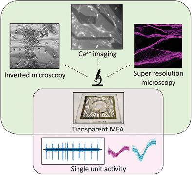
Advanced optical imaging techniques address important biological questions in neuroscience, where structures such as synapses are below the resolution limit of a conventional microscope. At the same time, microelectrode arrays (MEAs) are indispensable in understanding the language of neurons. Here, the authors show transparent MEAs capable of recording action potentials from neurons and compatible with advanced microscopy. The electrodes are made of the conducting polymer poly(3,4-ethylenedioxythiophene) doped with polystyrene sulfonate (PEDOT:PSS) and are patterned by optical lithography, ensuring scalable fabrication with good control over device parameters. A thickness of 380 nm ensures low enough impedance and >75% transparency throughout the visible part of the spectrum making them suitable for artefact-free recording in the presence of laser illumination. Using primary neuronal cells, the arrays record single units from multiple nearby sources with a signal-to-noise ratio of 7.7 (17.7 dB). Additionally, it is possible to perform calcium (Ca2+) imaging, a measure of neuronal activity, using the novel transparent electrodes. Different biomarkers are imaged through the electrodes using conventional and super-resolution microscopy (SRM), showing no qualitative differences compared to glass substrates. These transparent MEAs pave the way for harnessing the synergy between the superior temporal resolution of electrophysiology and the selectivity and high spatial resolution of optical imaging.
中文翻译:

用于同步电生理学和先进光学显微镜的微电极阵列
先进的光学成像技术解决了神经科学中的重要生物学问题,其中突触等结构低于传统显微镜的分辨率极限。同时,微电极阵列(MEA)对于理解神经元的语言是不可或缺的。在这里,作者展示了能够记录神经元动作电位并与先进显微镜兼容的透明 MEA。该电极由掺杂有聚苯乙烯磺酸盐(PEDOT:PSS)的导电聚合物聚(3,4-乙撑二氧噻吩)制成,并通过光学光刻进行图案化,确保可扩展制造并良好控制器件参数。 380 nm 的厚度可确保整个光谱的可见部分具有足够低的阻抗和 >75% 透明度,使其适合在激光照明下进行无伪影记录。该阵列使用原代神经元细胞,记录来自多个附近源的单个单元,信噪比为 7.7 (17.7 dB)。此外,还可以使用新型透明电极进行钙(Ca 2+ )成像,这是一种神经元活动的测量方法。使用传统和超分辨率显微镜(SRM)通过电极对不同的生物标记物进行成像,与玻璃基板相比没有显示出质量差异。这些透明的 MEA 为利用电生理学的卓越时间分辨率与光学成像的选择性和高空间分辨率之间的协同作用铺平了道路。
更新日期:2021-07-07
中文翻译:
用于同步电生理学和先进光学显微镜的微电极阵列
先进的光学成像技术解决了神经科学中的重要生物学问题,其中突触等结构低于传统显微镜的分辨率极限。同时,微电极阵列(MEA)对于理解神经元的语言是不可或缺的。在这里,作者展示了能够记录神经元动作电位并与先进显微镜兼容的透明 MEA。该电极由掺杂有聚苯乙烯磺酸盐(PEDOT:PSS)的导电聚合物聚(3,4-乙撑二氧噻吩)制成,并通过光学光刻进行图案化,确保可扩展制造并良好控制器件参数。 380 nm 的厚度可确保整个光谱的可见部分具有足够低的阻抗和 >75% 透明度,使其适合在激光照明下进行无伪影记录。该阵列使用原代神经元细胞,记录来自多个附近源的单个单元,信噪比为 7.7 (17.7 dB)。此外,还可以使用新型透明电极进行钙(Ca 2+ )成像,这是一种神经元活动的测量方法。使用传统和超分辨率显微镜(SRM)通过电极对不同的生物标记物进行成像,与玻璃基板相比没有显示出质量差异。这些透明的 MEA 为利用电生理学的卓越时间分辨率与光学成像的选择性和高空间分辨率之间的协同作用铺平了道路。


 京公网安备 11010802027423号