Journal of Structural Biology ( IF 3.0 ) Pub Date : 2021-05-08 , DOI: 10.1016/j.jsb.2021.107743 Sebastian Tacke 1 , Philipp Erdmann 2 , Zhexin Wang 1 , Sven Klumpe 2 , Michael Grange 1 , Jürgen Plitzko 2 , Stefan Raunser 1
|
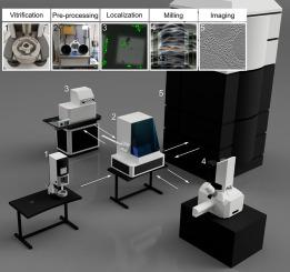
Cryo-electron tomography (cryo-ET) is an emerging technique to study the cellular architecture and the structure of proteins at high resolution in situ. Most biological specimens are too thick to be directly investigated and are therefore thinned by milling with a focused ion beam under cryogenic conditions (cryo-FIB). This procedure is prone to contaminations, which makes it a tedious process, often leading to suboptimal results. Here, we present new hardware that overcomes the current limitations. We developed a new glove box and a high vacuum cryo transfer system and installed a stage heater, a cryo-shield and a cryo-shutter in the FIB milling microscope. This reduces the ice contamination during the transfer and milling process and simplifies the handling of the sample. In addition, we tested a new software application that automates the key milling steps. Together, these improvements allow for high-quality, high-throughput cryo-FIB milling. This paves the way for new types of experiments, which have been previously considered infeasible.
中文翻译:
用于自动低温聚焦离子束铣削的简化工作流程
冷冻电子断层扫描 (cryo-ET) 是一种新兴的技术,可用于原位高分辨率研究细胞结构和蛋白质结构. 大多数生物标本太厚而无法直接研究,因此通过在低温条件下用聚焦离子束铣削 (cryo-FIB) 使其变薄。这个过程很容易受到污染,这使它成为一个乏味的过程,通常会导致不理想的结果。在这里,我们展示了克服当前限制的新硬件。我们开发了一个新的手套箱和一个高真空低温传输系统,并在 FIB 铣削显微镜中安装了一个载物台加热器、一个低温屏蔽和一个低温快门。这减少了转移和研磨过程中的冰污染,并简化了样品的处理。此外,我们测试了一个新的软件应用程序,它可以自动执行关键的铣削步骤。总之,这些改进可以实现高质量、高通量的冷冻 FIB 铣削。这为新型实验铺平了道路,


 京公网安备 11010802027423号