当前位置:
X-MOL 学术
›
Brain Pathol.
›
论文详情
Our official English website, www.x-mol.net, welcomes your feedback! (Note: you will need to create a separate account there.)
Diverse changes in microglia morphology and axonal pathology during the course of 1 year after mild traumatic brain injury in pigs
Brain Pathology ( IF 6.4 ) Pub Date : 2021-05-07 , DOI: 10.1111/bpa.12953 Michael R Grovola 1, 2 , Nicholas Paleologos 1, 2 , Daniel P Brown 1, 2 , Nathan Tran 1 , Kathryn L Wofford 1, 2 , James P Harris 1, 2 , Kevin D Browne 1, 2 , Patricia A Shewokis 3, 4 , John A Wolf 1, 2 , D Kacy Cullen 1, 2, 5 , John E Duda 1, 6, 7
Brain Pathology ( IF 6.4 ) Pub Date : 2021-05-07 , DOI: 10.1111/bpa.12953 Michael R Grovola 1, 2 , Nicholas Paleologos 1, 2 , Daniel P Brown 1, 2 , Nathan Tran 1 , Kathryn L Wofford 1, 2 , James P Harris 1, 2 , Kevin D Browne 1, 2 , Patricia A Shewokis 3, 4 , John A Wolf 1, 2 , D Kacy Cullen 1, 2, 5 , John E Duda 1, 6, 7
Affiliation
|
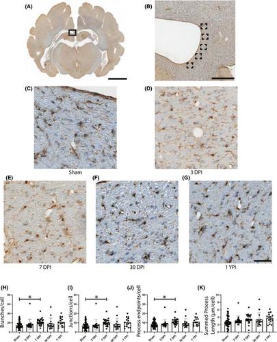
Over 2.8 million people experience mild traumatic brain injury (TBI) in the United States each year, which may lead to long-term neurological dysfunction. The mechanical forces that are caused by TBI propagate through the brain to produce diffuse axonal injury (DAI) and trigger secondary neuroinflammatory cascades. The cascades may persist from acute to chronic time points after injury, altering the homeostasis of the brain. However, the relationship between the hallmark axonal pathology of diffuse TBI and potential changes in glial cell activation or morphology have not been established in a clinically relevant large animal model at chronic time points. In this study, we assessed the tissue from pigs subjected to rapid head rotation in the coronal plane to generate mild TBI. Neuropathological assessments for axonal pathology, microglial morphological changes, and astrocyte reactivity were conducted in specimens out to 1-year post-injury. We detected an increase in overall amyloid precursor protein pathology, as well as periventricular white matter and fimbria/fornix pathology after a single mild TBI. We did not detect the changes in corpus callosum integrity or astrocyte reactivity. However, detailed microglial skeletal analysis revealed changes in morphology, most notably increases in the number of microglial branches, junctions, and endpoints. These subtle changes were most evident in periventricular white matter and certain hippocampal subfields, and were observed out to 1-year post-injury in some cases. These ongoing morphological alterations suggest persistent change in neuroimmune homeostasis. Additional studies are needed to characterize the underlying molecular and neurophysiological alterations, as well as potential contributions to neurological deficits.
中文翻译:

猪轻度脑外伤后1年内小胶质细胞形态和轴突病理的多样化变化
在美国,每年有超过 280 万人遭受轻度创伤性脑损伤 (TBI),这可能导致长期神经功能障碍。TBI 引起的机械力通过大脑传播,产生弥漫性轴突损伤 (DAI) 并引发继发性神经炎症级联反应。损伤后,级联反应可能会从急性时间点持续到慢性时间点,从而改变大脑的稳态。然而,在慢性时间点的临床相关大型动物模型中,尚未建立弥漫性 TBI 的标志性轴突病理学与神经胶质细胞活化或形态的潜在变化之间的关系。在这项研究中,我们评估了在冠状面快速旋转头部以产生轻度 TBI 的猪的组织。对损伤后一年内的标本进行轴突病理学、小胶质细胞形态变化和星形胶质细胞反应性的神经病理学评估。在一次轻度 TBI 后,我们检测到整体淀粉样前体蛋白病理学以及脑室周围白质和穹窿/穹窿病理学有所增加。我们没有检测到胼胝体完整性或星形胶质细胞反应性的变化。然而,详细的小胶质细胞骨骼分析揭示了形态的变化,最显着的是小胶质细胞分支、连接处和端点数量的增加。这些微妙的变化在脑室周围白质和某些海马亚区最为明显,并且在某些病例中观察到受伤后 1 年。这些持续的形态变化表明神经免疫稳态的持续变化。需要更多的研究来表征潜在的分子和神经生理学改变,以及对神经缺陷的潜在影响。
更新日期:2021-05-07
中文翻译:
猪轻度脑外伤后1年内小胶质细胞形态和轴突病理的多样化变化
在美国,每年有超过 280 万人遭受轻度创伤性脑损伤 (TBI),这可能导致长期神经功能障碍。TBI 引起的机械力通过大脑传播,产生弥漫性轴突损伤 (DAI) 并引发继发性神经炎症级联反应。损伤后,级联反应可能会从急性时间点持续到慢性时间点,从而改变大脑的稳态。然而,在慢性时间点的临床相关大型动物模型中,尚未建立弥漫性 TBI 的标志性轴突病理学与神经胶质细胞活化或形态的潜在变化之间的关系。在这项研究中,我们评估了在冠状面快速旋转头部以产生轻度 TBI 的猪的组织。对损伤后一年内的标本进行轴突病理学、小胶质细胞形态变化和星形胶质细胞反应性的神经病理学评估。在一次轻度 TBI 后,我们检测到整体淀粉样前体蛋白病理学以及脑室周围白质和穹窿/穹窿病理学有所增加。我们没有检测到胼胝体完整性或星形胶质细胞反应性的变化。然而,详细的小胶质细胞骨骼分析揭示了形态的变化,最显着的是小胶质细胞分支、连接处和端点数量的增加。这些微妙的变化在脑室周围白质和某些海马亚区最为明显,并且在某些病例中观察到受伤后 1 年。这些持续的形态变化表明神经免疫稳态的持续变化。需要更多的研究来表征潜在的分子和神经生理学改变,以及对神经缺陷的潜在影响。


 京公网安备 11010802027423号