当前位置:
X-MOL 学术
›
Stem Cells Transl. Med.
›
论文详情
Our official English website, www.x-mol.net, welcomes your
feedback! (Note: you will need to create a separate account there.)
Aging modulates the effects of ischemic injury upon mesenchymal cells within the renal interstitium and microvasculature
STEM CELLS Translational Medicine ( IF 5.4 ) Pub Date : 2021-05-05 , DOI: 10.1002/sctm.20-0392 Isaac W Shaw 1, 2 , Eoin D O'Sullivan 1, 3 , Angela O Pisco 4 , Gary Borthwick 1 , Kevin M Gallagher 1, 3 , Bruno Péault 2, 5 , Jeremy Hughes 1, 3 , David A Ferenbach 1, 3
STEM CELLS Translational Medicine ( IF 5.4 ) Pub Date : 2021-05-05 , DOI: 10.1002/sctm.20-0392 Isaac W Shaw 1, 2 , Eoin D O'Sullivan 1, 3 , Angela O Pisco 4 , Gary Borthwick 1 , Kevin M Gallagher 1, 3 , Bruno Péault 2, 5 , Jeremy Hughes 1, 3 , David A Ferenbach 1, 3
Affiliation
|
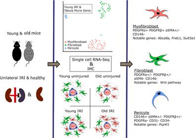
The renal mesenchyme contains heterogeneous cells, including interstitial fibroblasts and pericytes, with key roles in wound healing. Although healing is impaired in aged kidneys, the effect of age and injury on the mesenchyme remains poorly understood. We characterized renal mesenchymal cell heterogeneity in young vs old animals and after ischemia-reperfusion-injury (IRI) using multiplex immunolabeling and single cell transcriptomics. Expression patterns of perivascular cell markers (α-SMA, CD146, NG2, PDGFR-α, and PDGFR-β) correlated with their interstitial location. PDGFR-α and PDGFR-β co-expression labeled renal myofibroblasts more efficiently than the current standard marker α-SMA, and CD146 was a superior murine renal pericyte marker. Three renal mesenchymal subtypes; pericytes, fibroblasts, and myofibroblasts, were recapitulated with data from two independently performed single cell transcriptomic analyzes of murine kidneys, the first dataset an aging cohort and the second dataset injured kidneys following IRI. Mesenchymal cells segregated into subtypes with distinct patterns of expression with aging and following injury. Baseline uninjured old kidneys resembled post-ischemic young kidneys, with this phenotype further exaggerated following IRI. These studies demonstrate that age modulates renal perivascular/interstitial cell marker expression and transcriptome at baseline and in response to injury and provide tools for the histological and transcriptomic analysis of renal mesenchymal cells, paving the way for more accurate classification of renal mesenchymal cell heterogeneity and identification of age-specific pathways and targets.
中文翻译:

衰老调节缺血性损伤对肾间质和微血管内间充质细胞的影响
肾间充质含有异质细胞,包括间质成纤维细胞和周细胞,在伤口愈合中起关键作用。尽管老年肾脏的愈合受损,但年龄和损伤对间充质的影响仍然知之甚少。我们使用多重免疫标记和单细胞转录组学表征了年轻与老年动物和缺血再灌注损伤 (IRI) 后肾间充质细胞的异质性。血管周围细胞标志物(α-SMA、CD146、NG2、PDGFR-α 和 PDGFR-β)的表达模式与其间质位置相关。PDGFR-α 和 PDGFR-β 共表达标记肾肌成纤维细胞比目前的标准标记物 α-SMA 更有效,并且 CD146 是一种优越的鼠肾周细胞标记物。三种肾间充质亚型;周细胞、成纤维细胞和肌成纤维细胞,用来自两个独立进行的小鼠肾脏单细胞转录组分析的数据进行了概括,第一个数据集是老龄化队列,第二个数据集是 IRI 后的肾脏损伤。间充质细胞随着衰老和损伤而分离成具有不同表达模式的亚型。基线未受伤的老年肾脏类似于缺血后的年轻肾脏,这种表型在 IRI 后进一步夸大。这些研究表明,年龄在基线和对损伤作出反应时会调节肾血管周围/间质细胞标志物的表达和转录组,并为肾间充质细胞的组织学和转录组学分析提供工具,为更准确地分类和鉴定肾间充质细胞异质性铺平道路特定年龄的途径和目标。
更新日期:2021-05-05
中文翻译:
衰老调节缺血性损伤对肾间质和微血管内间充质细胞的影响
肾间充质含有异质细胞,包括间质成纤维细胞和周细胞,在伤口愈合中起关键作用。尽管老年肾脏的愈合受损,但年龄和损伤对间充质的影响仍然知之甚少。我们使用多重免疫标记和单细胞转录组学表征了年轻与老年动物和缺血再灌注损伤 (IRI) 后肾间充质细胞的异质性。血管周围细胞标志物(α-SMA、CD146、NG2、PDGFR-α 和 PDGFR-β)的表达模式与其间质位置相关。PDGFR-α 和 PDGFR-β 共表达标记肾肌成纤维细胞比目前的标准标记物 α-SMA 更有效,并且 CD146 是一种优越的鼠肾周细胞标记物。三种肾间充质亚型;周细胞、成纤维细胞和肌成纤维细胞,用来自两个独立进行的小鼠肾脏单细胞转录组分析的数据进行了概括,第一个数据集是老龄化队列,第二个数据集是 IRI 后的肾脏损伤。间充质细胞随着衰老和损伤而分离成具有不同表达模式的亚型。基线未受伤的老年肾脏类似于缺血后的年轻肾脏,这种表型在 IRI 后进一步夸大。这些研究表明,年龄在基线和对损伤作出反应时会调节肾血管周围/间质细胞标志物的表达和转录组,并为肾间充质细胞的组织学和转录组学分析提供工具,为更准确地分类和鉴定肾间充质细胞异质性铺平道路特定年龄的途径和目标。


 京公网安备 11010802027423号