Nanomedicine: Nanotechnology, Biology and Medicine ( IF 4.2 ) Pub Date : 2021-04-29 , DOI: 10.1016/j.nano.2021.102404 Hassan Rammal 1 , Almar Al Assaad 1 , Franco Dosio 2 , Barbara Stella 2 , Andrei Maksimenko 3 , Simona Mura 3 , Laurence Van Gulick 4 , Maïté Callewaert 5 , Didier Desmaële 3 , Patrick Couvreur 3 , Hamid Morjani 1 , Abdelilah Beljebbar 1
|
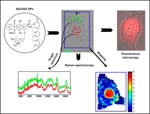
Intracellular distribution of doxorubicin (DOX) and its squalenoylated (SQ-DOX) nanoparticles (NPs) form in murine lung carcinoma M109 and human breast carcinoma MDA-MB-231 cells was investigated by Raman microspectroscopy. Pharmacological data showed that DOX induced higher cytotoxic effect than SQ-DOX NPs. Raman data were obtained using single-point measurements and imaging on the whole cell areas. These data showed that after DOX treatment at 1 μM, the spectral features of DOX were not detected in the M109 cell cytoplasm and nucleus. However, the intracellular distribution of SQ-DOX NPs was higher than DOX in the same conditions. In addition, SQ-DOX NPs were localized into both cell cytoplasm and nucleus. After 5 μM treatment, Raman bands of DOX at 1211 and 1241 cm−1 were detected in the nucleus. Moreover, the intensity ratio of these bands decreased, indicating DOX intercalation into DNA. However, after treatment with SQ-DOX NPs, the intensity of these Raman bands increased. Interestingly, with SQ-DOX NPs, the intensity of 1210/1241 cm−1 ratio was higher suggesting a lower fraction of intercalated DOX in DNA and higher amount of non-hydrolyzed SQ-DOX. Raman imaging data confirm this subcellular localization of these drugs in both M109 and MDA-MB-231 cells. These finding brings new insights to the cellular characterization of anticancer drugs at the molecular level, particularly in the field of nanomedicine.
中文翻译:
使用拉曼显微光谱研究角鲨烯-阿霉素在单个癌细胞内的分布和相互作用
通过拉曼显微光谱研究了多柔比星 (DOX) 及其角鲨烯酰化 (SQ-DOX) 纳米颗粒 (NPs) 在小鼠肺癌 M109 和人乳腺癌 MDA-MB-231 细胞中的细胞内分布。药理学数据表明,DOX 诱导的细胞毒作用高于 SQ-DOX NPs。拉曼数据是使用单点测量和整个细胞区域的成像获得的。这些数据表明,在 1 μM DOX 处理后,在 M109 细胞的细胞质和细胞核中未检测到 DOX 的光谱特征。然而,在相同条件下,SQ-DOX NPs 的细胞内分布高于 DOX。此外,SQ-DOX NPs 定位于细胞质和细胞核中。5 μM 处理后,DOX 在 1211 和 1241 cm -1的拉曼谱带在细胞核中检测到。此外,这些条带的强度比降低,表明 DOX 嵌入 DNA。然而,在用 SQ-DOX NPs 处理后,这些拉曼谱带的强度增加了。有趣的是,对于 SQ-DOX NPs,1210/1241 cm -1比率的强度更高,表明 DNA 中插入的 DOX 分数较低,未水解的 SQ-DOX 的量较高。拉曼成像数据证实了这些药物在 M109 和 MDA-MB-231 细胞中的这种亚细胞定位。这些发现为分子水平的抗癌药物的细胞表征带来了新的见解,特别是在纳米医学领域。


 京公网安备 11010802027423号