当前位置:
X-MOL 学术
›
Microsc. Res. Tech.
›
论文详情
Our official English website, www.x-mol.net, welcomes your
feedback! (Note: you will need to create a separate account there.)
Chemical and morphological changes of femtosecond laser-irradiated enamel using subablative parameters
Microscopy Research and Technique ( IF 2.0 ) Pub Date : 2021-04-27 , DOI: 10.1002/jemt.23795 Heitor Hussni Casarin 1 , Vicente Silva Mattos 2 , Jarbas Caiado de Castro Neto 2 , Michelle Alexandra Chinelatti 1
Microscopy Research and Technique ( IF 2.0 ) Pub Date : 2021-04-27 , DOI: 10.1002/jemt.23795 Heitor Hussni Casarin 1 , Vicente Silva Mattos 2 , Jarbas Caiado de Castro Neto 2 , Michelle Alexandra Chinelatti 1
Affiliation
|
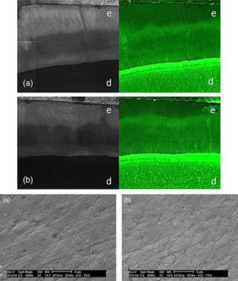
Chemical composition of dental enamel has a great relationship with the prevention of caries. The objective of the present work was to evaluate the chemical and morphological changes of femtosecond laser-irradiated enamel with subablative parameters using Raman spectroscopy, confocal laser scanning microscopy (CLSM), and scanning electron microscopy (SEM). Bovine incisor teeth were used to obtain 30 enamel specimens (5 × 5 mm2). The chemical composition of the control sample was analyzed by Raman spectrometry to acquire the absorption spectrum, delimiting the areas under the carbonate and phosphate bands. This analysis was used to evaluate the change in the chemical composition of the sample after irradiation. The specimens were irradiated (IRR) with a Ti:Sapphire laser system (pulsed and focused modes, femtosecond regime 70 fs, average power of 1 W and exposure time of 15 s). After irradiation, the areas under the carbonate and phosphate absorption bands were delimited in each specimen. Raman spectrometry data were analyzed using Student's t-test (α = 5%). By comparing the spectra of the IRR and non-irradiated (NI) specimens, the results showed a significant increase in the area value for the phosphate peaks and a significant reduction in the area value for the carbonate peak and the carbonate:phosphate ratio. CLSM and SEM analyses did not reveal structural alterations in the subsurface nor morphological alterations in the IRR enamel surface, respectively. It was concluded that femtosecond laser irradiation using subablative parameters reduced the carbonate content and the carbonate/phosphate ratio without altering the structure and morphology of the dental enamel.
中文翻译:

使用亚烧蚀参数的飞秒激光辐照牙釉质的化学和形态变化
牙釉质的化学成分与预防龋齿有很大关系。本工作的目的是使用拉曼光谱、共聚焦激光扫描显微镜 (CLSM) 和扫描电子显微镜 (SEM) 评估具有亚烧蚀参数的飞秒激光照射牙釉质的化学和形态变化。使用牛门牙获得 30 个牙釉质标本(5 × 5 mm 2)。通过拉曼光谱法分析对照样品的化学成分以获得吸收光谱,划定碳酸盐和磷酸盐带下的区域。该分析用于评估辐照后样品化学成分的变化。用钛:蓝宝石激光系统(脉冲和聚焦模式,飞秒状态 70 fs,1 W 平均功率和 15 s 曝光时间)对样品进行辐照 (IRR)。辐照后,在每个样品中划定碳酸盐和磷酸盐吸收带下的区域。使用Student's t分析拉曼光谱数据- 测试(α = 5%)。通过比较 IRR 和未辐照 (NI) 样品的光谱,结果显示磷酸盐峰的面积值显着增加,而碳酸盐峰的面积值和碳酸盐:磷酸盐比值显着降低。CLSM 和 SEM 分析分别没有显示下表面的结构改变和 IRR 牙釉质表面的形态改变。得出的结论是,使用亚烧蚀参数的飞秒激光照射降低了碳酸盐含量和碳酸盐/磷酸盐比率,而不会改变牙釉质的结构和形态。
更新日期:2021-04-27
中文翻译:
使用亚烧蚀参数的飞秒激光辐照牙釉质的化学和形态变化
牙釉质的化学成分与预防龋齿有很大关系。本工作的目的是使用拉曼光谱、共聚焦激光扫描显微镜 (CLSM) 和扫描电子显微镜 (SEM) 评估具有亚烧蚀参数的飞秒激光照射牙釉质的化学和形态变化。使用牛门牙获得 30 个牙釉质标本(5 × 5 mm 2)。通过拉曼光谱法分析对照样品的化学成分以获得吸收光谱,划定碳酸盐和磷酸盐带下的区域。该分析用于评估辐照后样品化学成分的变化。用钛:蓝宝石激光系统(脉冲和聚焦模式,飞秒状态 70 fs,1 W 平均功率和 15 s 曝光时间)对样品进行辐照 (IRR)。辐照后,在每个样品中划定碳酸盐和磷酸盐吸收带下的区域。使用Student's t分析拉曼光谱数据- 测试(α = 5%)。通过比较 IRR 和未辐照 (NI) 样品的光谱,结果显示磷酸盐峰的面积值显着增加,而碳酸盐峰的面积值和碳酸盐:磷酸盐比值显着降低。CLSM 和 SEM 分析分别没有显示下表面的结构改变和 IRR 牙釉质表面的形态改变。得出的结论是,使用亚烧蚀参数的飞秒激光照射降低了碳酸盐含量和碳酸盐/磷酸盐比率,而不会改变牙釉质的结构和形态。


 京公网安备 11010802027423号