当前位置:
X-MOL 学术
›
Stem Cells Transl. Med.
›
论文详情
Our official English website, www.x-mol.net, welcomes your
feedback! (Note: you will need to create a separate account there.)
Mesenchymal stem cells and three-dimensional-osteoconductive scaffold regenerate calvarial bone in critical size defects in swine
STEM CELLS Translational Medicine ( IF 5.4 ) Pub Date : 2021-04-01 , DOI: 10.1002/sctm.20-0534 Zoe M Johnson 1 , Yuan Yuan 1 , Xiangjia Li 2, 3 , Tea Jashashvili 4 , Michael Jamieson 5 , Mark Urata 6 , Yong Chen 3 , Yang Chai 1
STEM CELLS Translational Medicine ( IF 5.4 ) Pub Date : 2021-04-01 , DOI: 10.1002/sctm.20-0534 Zoe M Johnson 1 , Yuan Yuan 1 , Xiangjia Li 2, 3 , Tea Jashashvili 4 , Michael Jamieson 5 , Mark Urata 6 , Yong Chen 3 , Yang Chai 1
Affiliation
|
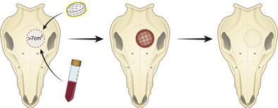
Craniofacial bones protect vital organs, perform important physiological functions, and shape facial identity. Critical-size defects (CSDs) in calvarial bones, which will not heal spontaneously, are caused by trauma, congenital defects, or tumor resections. They pose a great challenge for patients and physicians, and significantly compromise quality of life. Currently, calvarial CSDs are treated either by allogenic or autologous grafts, metal or other synthetic plates that are associated with considerable complications. While previous studies have explored tissue regeneration for calvarial defects, most have been done in small animal models with limited translational value. Here we define a swine calvarial CSD model and show a novel approach to regenerate high-quality bone in these defects by combining mesenchymal stem cells (MSCs) with a three-dimensional (3D)-printed osteoconductive HA/TCP scaffold. Specifically, we have compared the performance of dental pulp neural crest MSCs (DPNCCs) to bone marrow aspirate (BMA) combined with a 3D-printed HA/TCP scaffold to regenerate bone in a calvarial CSD (>7.0 cm2). Both DPNCCs and BMA loaded onto the 3D-printed osteoconductive scaffold support the regeneration of calvarial bone with density, compression strength, and trabecular structures similar to native bone. Our study demonstrates a novel application of an original scaffold design combined with DPNCCs or BMA to support regeneration of high-quality bone in a newly defined and clinically relevant swine calvarial CSD model. This discovery may have important impact on bone regeneration beyond the craniofacial region and will ultimately benefit patients who suffer from debilitating CSDs.
中文翻译:

间充质干细胞和三维骨传导支架在猪的临界尺寸缺陷中再生颅骨
颅面骨骼保护重要器官,执行重要的生理功能,并塑造面部特征。颅骨中的临界尺寸缺陷 (CSD) 不会自发愈合,是由创伤、先天性缺陷或肿瘤切除引起的。它们对患者和医生构成了巨大挑战,并严重影响了生活质量。目前,颅骨 CSD 通过同种异体或自体移植物、金属或其他与相当多的并发症相关的合成板进行治疗。虽然以前的研究已经探索了颅骨缺损的组织再生,但大多数是在具有有限转化价值的小动物模型中完成的。在这里,我们定义了一个猪颅骨 CSD 模型,并展示了一种通过将间充质干细胞 (MSCs) 与三维 (3D) 打印的骨传导性 HA/TCP 支架相结合来在这些缺陷中再生高质量骨骼的新方法。具体来说,我们比较了牙髓神经嵴 MSCs (DPNCCs) 与骨髓抽吸物 (BMA) 结合 3D 打印 HA/TCP 支架在颅骨 CSD (>7.0 cm) 中再生骨的性能2 )。加载到 3D 打印的骨传导支架上的 DPNCC 和 BMA 都支持颅骨的再生,其密度、压缩强度和与天然骨相似的小梁结构。我们的研究展示了原始支架设计与 DPNCC 或 BMA 相结合的新应用,以支持新定义的和临床相关的猪颅骨 CSD 模型中高质量骨骼的再生。这一发现可能对颅面区域以外的骨再生产生重要影响,最终将使患有衰弱性 CSD 的患者受益。
更新日期:2021-04-01
中文翻译:
间充质干细胞和三维骨传导支架在猪的临界尺寸缺陷中再生颅骨
颅面骨骼保护重要器官,执行重要的生理功能,并塑造面部特征。颅骨中的临界尺寸缺陷 (CSD) 不会自发愈合,是由创伤、先天性缺陷或肿瘤切除引起的。它们对患者和医生构成了巨大挑战,并严重影响了生活质量。目前,颅骨 CSD 通过同种异体或自体移植物、金属或其他与相当多的并发症相关的合成板进行治疗。虽然以前的研究已经探索了颅骨缺损的组织再生,但大多数是在具有有限转化价值的小动物模型中完成的。在这里,我们定义了一个猪颅骨 CSD 模型,并展示了一种通过将间充质干细胞 (MSCs) 与三维 (3D) 打印的骨传导性 HA/TCP 支架相结合来在这些缺陷中再生高质量骨骼的新方法。具体来说,我们比较了牙髓神经嵴 MSCs (DPNCCs) 与骨髓抽吸物 (BMA) 结合 3D 打印 HA/TCP 支架在颅骨 CSD (>7.0 cm) 中再生骨的性能2 )。加载到 3D 打印的骨传导支架上的 DPNCC 和 BMA 都支持颅骨的再生,其密度、压缩强度和与天然骨相似的小梁结构。我们的研究展示了原始支架设计与 DPNCC 或 BMA 相结合的新应用,以支持新定义的和临床相关的猪颅骨 CSD 模型中高质量骨骼的再生。这一发现可能对颅面区域以外的骨再生产生重要影响,最终将使患有衰弱性 CSD 的患者受益。


 京公网安备 11010802027423号