当前位置:
X-MOL 学术
›
J. Appl. Crystallogr.
›
论文详情
Our official English website, www.x-mol.net, welcomes your
feedback! (Note: you will need to create a separate account there.)
2D real space visualization of d values in polycrystalline bulk materials of different hardness
Journal of Applied Crystallography ( IF 5.2 ) Pub Date : 2021-03-31 , DOI: 10.1107/s1600576721001631 Mari Mizusawa , Kenji Sakurai
Journal of Applied Crystallography ( IF 5.2 ) Pub Date : 2021-03-31 , DOI: 10.1107/s1600576721001631 Mari Mizusawa , Kenji Sakurai
|
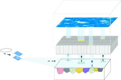
Conventional X‐ray diffraction measurements provide some average structural information, mainly on the crystal structure of the whole area of the given specimen, which might not be very uniform and may include different crystal structures, such as co‐existing crystal phases and/or lattice distortion. The way in which the lattice plane changes due to strain also might depend on the position in the sample, and the average information might have some limits. Therefore, it is important to analyse the sample with good lateral spatial resolution in real space. Although various techniques for diffraction topography have been developed for single crystals, it has not always been easy to image polycrystalline materials. Since the late 1990s, imaging technology for fluorescent X‐rays and X‐ray absorption fine structure has been developed via a method that does not scan either a sample or an X‐ray beam. X‐ray diffraction imaging can be performed when this technique is applied to a synchrotron radiation beamline with a variable wavelength. The present paper reports the application of X‐ray diffraction imaging to bulk steel materials with varying hardness. In this study, the distribution of lattice distortion of hardness test blocks with different hardness was examined. Via this 2D visualization method, the grains of the crystals with low hardness are large enough to be observed by X‐ray diffraction contrast in real space. The change of the d value in the vicinity of the Vickers mark has also been quantitatively evaluated.
中文翻译:

不同硬度的多晶块状材料中d值的二维真实空间可视化
常规X射线衍射测量可提供一些平均结构信息,主要是有关给定样品整个区域的晶体结构,这些信息可能不是很均匀,并且可能包含不同的晶体结构,例如共存的晶体相和/或晶格失真。晶格平面因应变而变化的方式也可能取决于样品中的位置,并且平均信息可能会有一些限制。因此,重要的是要在实际空间中分析具有良好横向空间分辨率的样本。尽管已经针对单晶开发了各种用于衍射形貌的技术,但是对多晶材料进行成像并不总是容易的。自1990年代后期以来,荧光X射线和X射线吸收精细结构的成像技术是通过不扫描样品或X射线束的方法开发的。当将此技术应用于具有可变波长的同步辐射束线时,可以执行X射线衍射成像。本文报道了X射线衍射成像技术在不同硬度的散装钢材上的应用。在这项研究中,检查了具有不同硬度的硬度测试块的晶格畸变分布。通过这种2D可视化方法,低硬度晶体的晶粒足够大,可以在实际空间中通过X射线衍射对比观察到。的变化 当将此技术应用于具有可变波长的同步辐射束线时,可以执行X射线衍射成像。本文报道了X射线衍射成像技术在不同硬度的散装钢材上的应用。在这项研究中,检查了具有不同硬度的硬度测试块的晶格畸变分布。通过这种2D可视化方法,低硬度晶体的晶粒足够大,可以在实际空间中通过X射线衍射对比观察到。的变化 当将此技术应用于具有可变波长的同步辐射束线时,可以执行X射线衍射成像。本文报道了X射线衍射成像技术在不同硬度的散装钢材上的应用。在这项研究中,检查了具有不同硬度的硬度测试块的晶格畸变分布。通过这种2D可视化方法,低硬度晶体的晶粒足够大,可以在实际空间中通过X射线衍射对比观察到。的变化 低硬度晶体的晶粒足够大,可以在实际空间中通过X射线衍射对比观察到。的变化 低硬度晶体的晶粒足够大,可以在实际空间中通过X射线衍射对比观察到。的变化维克斯标记附近的d值也已进行了定量评估。
更新日期:2021-04-06
中文翻译:
不同硬度的多晶块状材料中d值的二维真实空间可视化
常规X射线衍射测量可提供一些平均结构信息,主要是有关给定样品整个区域的晶体结构,这些信息可能不是很均匀,并且可能包含不同的晶体结构,例如共存的晶体相和/或晶格失真。晶格平面因应变而变化的方式也可能取决于样品中的位置,并且平均信息可能会有一些限制。因此,重要的是要在实际空间中分析具有良好横向空间分辨率的样本。尽管已经针对单晶开发了各种用于衍射形貌的技术,但是对多晶材料进行成像并不总是容易的。自1990年代后期以来,荧光X射线和X射线吸收精细结构的成像技术是通过不扫描样品或X射线束的方法开发的。当将此技术应用于具有可变波长的同步辐射束线时,可以执行X射线衍射成像。本文报道了X射线衍射成像技术在不同硬度的散装钢材上的应用。在这项研究中,检查了具有不同硬度的硬度测试块的晶格畸变分布。通过这种2D可视化方法,低硬度晶体的晶粒足够大,可以在实际空间中通过X射线衍射对比观察到。的变化 当将此技术应用于具有可变波长的同步辐射束线时,可以执行X射线衍射成像。本文报道了X射线衍射成像技术在不同硬度的散装钢材上的应用。在这项研究中,检查了具有不同硬度的硬度测试块的晶格畸变分布。通过这种2D可视化方法,低硬度晶体的晶粒足够大,可以在实际空间中通过X射线衍射对比观察到。的变化 当将此技术应用于具有可变波长的同步辐射束线时,可以执行X射线衍射成像。本文报道了X射线衍射成像技术在不同硬度的散装钢材上的应用。在这项研究中,检查了具有不同硬度的硬度测试块的晶格畸变分布。通过这种2D可视化方法,低硬度晶体的晶粒足够大,可以在实际空间中通过X射线衍射对比观察到。的变化 低硬度晶体的晶粒足够大,可以在实际空间中通过X射线衍射对比观察到。的变化 低硬度晶体的晶粒足够大,可以在实际空间中通过X射线衍射对比观察到。的变化维克斯标记附近的d值也已进行了定量评估。


 京公网安备 11010802027423号