Artificial Intelligence in Medicine ( IF 6.1 ) Pub Date : 2021-03-24 , DOI: 10.1016/j.artmed.2021.102057 Abbas Memiş 1 , Songül Varlı 1 , Fuat Bilgili 2
|
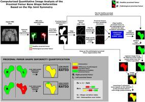
As a result of most of the bone disorders seen in hip joints, shape deformities occur in the structural form of the hip joint components. Image-based quantitative analysis and assessment of these deformities in bone shapes are very important for the evaluation, treatment, and prognosis of the various hip joint bone disorders. In this article, a novel approach for the image-based computerized quantitative analysis of proximal femur shape deformities is presented. In the proposed approach, shape deformities of the pathological proximal femurs were quantified over the contralateral healthy proximal femur shape structure of the same patient in 2D by taking the hip joint symmetry property of human anatomy into consideration. It is based on the idea that if the right and left proximal femurs in bilateral hip joints are highly symmetrical and also if one of the proximal femurs is healthy and the contralateral one is pathological, the non-overlapping bone shape regions can represent the deformities in pathological proximal femurs when both proximal femurs are registered to overlap each other. In the methodological process of the proposed study, a set of image preprocessing operations was primarily performed on the raw magnetic resonance imaging (MRI) data. Then, the segmented proximal femurs in bilateral hip joint images were automatically aligned with the Iterative Closest Point (ICP) rigid registration method. Following the registration, a set of image postprocessing operations was performed on the images of proximal femurs aligned. In the quantification phase, the bone shape deformities in pathological proximal femurs were quantified simply in terms of the mismatching area in 2D by measuring a shape variation index representing the total bone shape deformity ratio. To evaluate the proposed quantitative shape analysis approach, bilateral hip joints in a total of 13 coronal MRI sections of 13 patients with Legg-Calve-Perthes disease (LCPD) were used. Experimental studies have shown that the proposed approach has quite promising results in the quantitative representation of the pathological proximal femur shape deformities. Furthermore, consistent results have been observed for the Waldenström classification stages of the disease. The shape deformity ratios in pathological proximal femurs were quantified as 9.44% (1.40), 18.38% (6.30), 24.73% (12.42), and 27.66% (10.41), respectively for the Initial, Fragmentation, Reossification, and Remodelling stages of LCPD with the quantification error rates of 0.29% (0.16), 0.58% (0.71), 1.12% (0.82), and 0.80% (0.98). Additionally, a mean error rate of 0.65% (0.68) was observed for the quantified shape deformity ratios of all samples.
中文翻译:
基于髋关节对称性的股骨近端骨形状畸形计算机定量图像分析的新方法
由于髋关节中出现的大多数骨骼疾病,髋关节组件的结构形式会出现形状畸形。这些骨骼形状畸形的基于图像的定量分析和评估对于各种髋关节骨骼疾病的评估、治疗和预后非常重要。在本文中,提出了一种基于图像的股骨近端形状畸形计算机定量分析的新方法。在所提出的方法中,通过考虑人体解剖学的髋关节对称性,在 2D 中在同一患者的对侧健康股骨近端形状结构上量化病理性股骨近端的形状畸形。基于这样的想法,如果双侧髋关节的左右股骨近端高度对称,并且如果近端股骨之一是健康的,而对侧的股骨是病理性的,那么不重叠的骨形区域可以代表髋关节的畸形。当两个股骨近端都注册为彼此重叠时,病理性股骨近端。在拟议研究的方法论过程中,主要对原始磁共振成像 (MRI) 数据执行一组图像预处理操作。然后,将双侧髋关节图像中分割的股骨近端与迭代最近点(ICP)刚性配准方法自动对齐。配准后,对对齐的股骨近端图像执行一组图像后处理操作。在量化阶段,病理性股骨近端的骨形状畸形通过测量代表总骨形状畸形比率的形状变化指数,简单地根据 2D 中的不匹配区域进行量化。为了评估所提出的定量形状分析方法,使用了 13 名 Legg-Calve-Perthes 病 (LCPD) 患者的总共 13 个冠状 MRI 切片中的双侧髋关节。实验研究表明,所提出的方法在病理性股骨近端形状畸形的定量表示方面具有非常有希望的结果。此外,对于该疾病的 Waldenström 分类阶段,已经观察到一致的结果。病理性股骨近端形状畸形率量化为 9.44%(使用了 13 名 Legg-Calve-Perthes 病 (LCPD) 患者的 13 个冠状位 MRI 切片中的双侧髋关节。实验研究表明,所提出的方法在病理性股骨近端形状畸形的定量表示方面具有非常有希望的结果。此外,对于该疾病的 Waldenström 分类阶段,已经观察到一致的结果。病理性股骨近端形状畸形率量化为 9.44%(使用了 13 名 Legg-Calve-Perthes 病 (LCPD) 患者的 13 个冠状位 MRI 切片中的双侧髋关节。实验研究表明,所提出的方法在病理性股骨近端形状畸形的定量表示方面具有非常有希望的结果。此外,对于该疾病的 Waldenström 分类阶段,已经观察到一致的结果。病理性股骨近端形状畸形率量化为 9.44%(1.40), 18.38% (6.30), 24.73% (12.42) 和 27.66% (10.41),分别用于 LCPD 的初始、碎片化、再骨化和重塑阶段,量化错误率为 0.29% (0.16), 0.58% (0.71), 1.12% (0.82) 和 0.80% (0.98)。此外,平均错误率为 0.65% (0.68) 观察到所有样品的量化形状变形率。


 京公网安备 11010802027423号