当前位置:
X-MOL 学术
›
Brain Pathol.
›
论文详情
Our official English website, www.x-mol.net, welcomes your
feedback! (Note: you will need to create a separate account there.)
Region-specific preservation of Purkinje cell morphology and motor behavior in the ATXN1[82Q] mouse model of spinocerebellar ataxia 1
Brain Pathology ( IF 5.8 ) Pub Date : 2021-03-16 , DOI: 10.1111/bpa.12946 Joshua J White 1 , Laurens W J Bosman 1 , Francois G C Blot 1 , Catarina Osório 1 , Bram W Kuppens 1 , Wilhelmina H J J Krijnen 1 , Charlotte Andriessen 1 , Chris I De Zeeuw 1, 2 , Dick Jaarsma 1 , Martijn Schonewille 1
Brain Pathology ( IF 5.8 ) Pub Date : 2021-03-16 , DOI: 10.1111/bpa.12946 Joshua J White 1 , Laurens W J Bosman 1 , Francois G C Blot 1 , Catarina Osório 1 , Bram W Kuppens 1 , Wilhelmina H J J Krijnen 1 , Charlotte Andriessen 1 , Chris I De Zeeuw 1, 2 , Dick Jaarsma 1 , Martijn Schonewille 1
Affiliation
|
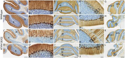
Purkinje cells are the primary processing units of the cerebellar cortex and display molecular heterogeneity that aligns with differences in physiological properties, projection patterns, and susceptibility to disease. In particular, multiple mouse models that feature Purkinje cell degeneration are characterized by incomplete and patterned Purkinje cell degeneration, suggestive of relative sparing of Purkinje cell subpopulations, such as those expressing Aldolase C/zebrinII (AldoC) or residing in the vestibulo-cerebellum. Here, we investigated a well-characterized Purkinje cell-specific mouse model for spinocerebellar ataxia type 1 (SCA1) that expresses human ATXN1 with a polyQ expansion (82Q). Our pathological analysis confirms previous findings that Purkinje cells of the vestibulo-cerebellum, i.e., the flocculonodular lobes, and crus I are relatively spared from key pathological hallmarks: somatodendritic atrophy, and the appearance of p62/SQSTM1-positive inclusions. However, immunohistological analysis of transgene expression revealed that spared Purkinje cells do not express mutant ATXN1 protein, indicating the sparing of Purkinje cells can be explained by an absence of transgene expression. Additionally, we found that Purkinje cells in other cerebellar lobules that typically express AldoC, not only display severe pathology but also show loss of AldoC expression. The relatively preserved flocculonodular lobes and crus I showed a substantial fraction of Purkinje cells that expressed the mutant protein and displayed pathology as well as loss of AldoC expression. Despite considerable pathology in these lobules, behavioral analyses demonstrated a relative sparing of related functions, suggestive of sufficient functional cerebellar reserve. Together, the data indicate that mutant ATXN1 affects both AldoC-positive and AldoC-negative Purkinje cells and disrupts normal parasagittal AldoC expression in Purkinje cells. Our results show that, in a mouse model otherwise characterized by widespread Purkinje cell degeneration, sparing of specific subpopulations is sufficient to maintain normal performance of specific behaviors within the context of the functional, modular map of the cerebellum.
中文翻译:

脊髓小脑共济失调1 ATXN1[82Q]小鼠模型中浦肯野细胞形态和运动行为的区域特异性保存
浦肯野细胞是小脑皮层的主要处理单元,其分子异质性与生理特性、投射模式和疾病易感性的差异一致。特别是,具有浦肯野细胞变性的多个小鼠模型的特点是不完全和模式化的浦肯野细胞变性,这表明浦肯野细胞亚群相对保留,例如表达醛缩酶 C/zebrinII (AldoC) 或位于前庭小脑中的那些。在这里,我们研究了一个充分表征的浦肯野细胞特异性小鼠模型,用于表达具有 polyQ 扩展 (82Q) 的人类 ATXN1 的脊髓小脑共济失调 1 型 (SCA1)。我们的病理学分析证实了先前的发现,即前庭小脑的浦肯野细胞,即绒球结节叶,和小腿 I 相对免受关键病理特征的影响:体树突萎缩和 p62/SQSTM1 阳性包涵体的出现。然而,转基因表达的免疫组织学分析显示,幸存的浦肯野细胞不表达突变的 ATXN1 蛋白,这表明浦肯野细胞的幸存可以通过没有转基因表达来解释。此外,我们发现其他小脑小叶中通常表达 AldoC 的浦肯野细胞不仅表现出严重的病理,而且表现出 AldoC 表达的丧失。相对保存的绒球结节叶和小腿 I 显示了相当一部分浦肯野细胞,它们表达了突变蛋白并表现出病理学以及 AldoC 表达的丧失。尽管这些小叶有相当大的病理学,行为分析表明相关功能相对保留,提示有足够的小脑功能储备。总之,数据表明突变 ATXN1 影响 AldoC 阳性和 AldoC 阴性浦肯野细胞,并破坏浦肯野细胞中正常的矢状旁 AldoC 表达。我们的研究结果表明,在以广泛的浦肯野细胞变性为特征的小鼠模型中,保留特定亚群足以在小脑的功能性模块化图谱的范围内维持特定行为的正常表现。
更新日期:2021-03-16
中文翻译:
脊髓小脑共济失调1 ATXN1[82Q]小鼠模型中浦肯野细胞形态和运动行为的区域特异性保存
浦肯野细胞是小脑皮层的主要处理单元,其分子异质性与生理特性、投射模式和疾病易感性的差异一致。特别是,具有浦肯野细胞变性的多个小鼠模型的特点是不完全和模式化的浦肯野细胞变性,这表明浦肯野细胞亚群相对保留,例如表达醛缩酶 C/zebrinII (AldoC) 或位于前庭小脑中的那些。在这里,我们研究了一个充分表征的浦肯野细胞特异性小鼠模型,用于表达具有 polyQ 扩展 (82Q) 的人类 ATXN1 的脊髓小脑共济失调 1 型 (SCA1)。我们的病理学分析证实了先前的发现,即前庭小脑的浦肯野细胞,即绒球结节叶,和小腿 I 相对免受关键病理特征的影响:体树突萎缩和 p62/SQSTM1 阳性包涵体的出现。然而,转基因表达的免疫组织学分析显示,幸存的浦肯野细胞不表达突变的 ATXN1 蛋白,这表明浦肯野细胞的幸存可以通过没有转基因表达来解释。此外,我们发现其他小脑小叶中通常表达 AldoC 的浦肯野细胞不仅表现出严重的病理,而且表现出 AldoC 表达的丧失。相对保存的绒球结节叶和小腿 I 显示了相当一部分浦肯野细胞,它们表达了突变蛋白并表现出病理学以及 AldoC 表达的丧失。尽管这些小叶有相当大的病理学,行为分析表明相关功能相对保留,提示有足够的小脑功能储备。总之,数据表明突变 ATXN1 影响 AldoC 阳性和 AldoC 阴性浦肯野细胞,并破坏浦肯野细胞中正常的矢状旁 AldoC 表达。我们的研究结果表明,在以广泛的浦肯野细胞变性为特征的小鼠模型中,保留特定亚群足以在小脑的功能性模块化图谱的范围内维持特定行为的正常表现。


 京公网安备 11010802027423号