Clinical Spectroscopy Pub Date : 2021-02-27 , DOI: 10.1016/j.clispe.2021.100006 Shachi Mittal , Tomasz P. Wrobel , Michael Walsh , Andre Kajdacsy-Balla , Rohit Bhargava
|
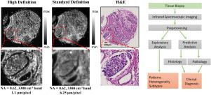
Digital analysis of cancer specimens using spectroscopic imaging coupled to machine learning is an emerging area that links spatially localized spectral signatures to tissue structure and disease. In this study, we examine the role of spatial-spectral tradeoffs in infrared spectroscopic imaging configurations for probing tumors and the associated microenvironment profiles at different levels of model complexity. We image breast tissue using standard and high-definition Fourier Transform Infrared (FT-IR) imaging and systematically examine the localization, spectral origins, and utility of data for classification. Results demonstrate that higher spatial detail provides high sensitivity and specificity of tissue segmentation, despite the increased subcellular variability. High definition imaging also allows accurate analysis of complex, multiclass models of breast tissue without compromising accuracy. A comparison of results also highlights the key differences in the data distributions and classification performance across modalities to better guide decision making for acquiring and analyzing IR imaging data for specific histopathological models.
中文翻译:
使用红外光谱成像的乳腺癌组织病理学:仪器配置的影响
使用结合机器学习的光谱成像对癌症标本进行数字分析是一个新兴的领域,它将空间定位的光谱特征与组织结构和疾病联系在一起。在这项研究中,我们检查了空间光谱权衡在探测光谱的红外光谱成像配置中的作用以及不同级别的模型复杂性相关的微环境配置文件。我们使用标准和高清傅立叶变换红外(FT-IR)成像对乳房组织进行成像,并系统地检查定位,光谱来源和分类数据的实用性。结果表明,尽管增加了亚细胞变异性,但较高的空间细节提供了组织分割的高灵敏度和特异性。高清晰成像还可以对复杂的,多种多样的乳腺组织模型,而不会影响准确性。结果的比较还突出显示了跨模式的数据分布和分类性能的关键差异,以更好地指导针对特定组织病理学模型获取和分析IR成像数据的决策。


 京公网安备 11010802027423号