Spectrochimica Acta Part A: Molecular and Biomolecular Spectroscopy ( IF 4.4 ) Pub Date : 2021-02-22 , DOI: 10.1016/j.saa.2021.119607 Fran Nekvapil , Alexander Bunge , Lucian Barbu Tudoran , Simona Cintă Pinzaru
|
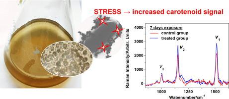
Ferrite nanoparticles are increasingly produced and exploited as adsorbents for environmental pollutants. However, their impact on the aquatic microbiota such as cyanobacteria, are not yet investigated. Targeting the environmental monitoring context, in this paper we explored for the first time if any change in the carotenoid signal from cyanobacteria Coelomoron pussilum (AICB 1012) exposed to non-lethal doses of Mn and Zn doped ferrite nanoparticles (NPs) can be associated with the nano-aggression on single-cell level, using micro-Raman spectroscopy. UV–Vis absorption spectroscopy of the bulk culture and single-cell Raman microscopy showed that the carotenoid signal increases relative to the chlorophyll contribution upon exposure of the cells to the Mn-ferrite NPs throughout the 7 days of the experiment. The red-shift and broadening of the strongest carotenoid Raman band arising from (C C) stretching modes indicates the change of carotenoid profile towards increased amount of β-carotene in answer to the NPs stress. The increase of this band intensity relative to the fluorescence background was also observed in Zn-ferrite NPs treatment. Using a simplified and rapid sample preparation procedure, electron microscopy in both transmission and scanning modes, showed greater coverage of the cells by the stable colloidal AgNPs than by the magnetic ferrite NPs. The latter mostly clumped together rather than adhering to the cells. The combined single-cell micro-Raman tracking of physiological response of the unicellular photosynthetic microorganisms coupled with electron microscopy approach to visualise cell-NPs interaction and the extracellular polymeric substance secretion holds the promise for rapid assessment of the NPs-induced environmental stress acting on the unicellular organisms.
中文翻译:
单细胞拉曼显微光谱法用于跟踪暴露于Mn和Zn掺杂的铁氧体纳米粒子的蓝细菌中的类胡萝卜素
铁氧体纳米颗粒的生产越来越多,并被用作环境污染物的吸附剂。然而,尚未研究它们对诸如蓝细菌的水生微生物的影响。针对环境监测环境,本文首次探讨了蓝藻腔肠龙的类胡萝卜素信号是否发生任何变化(AICB 1012)暴露于非致死剂量的Mn和Zn掺杂的铁氧体纳米颗粒(NPs)可以使用微拉曼光谱法与单细胞水平的纳米侵略性相关。整个培养和单细胞拉曼显微镜的紫外可见吸收光谱显示,在整个实验的7天中,当细胞暴露于锰铁氧体NP时,类胡萝卜素信号相对于叶绿素的贡献增加。由(CC)拉伸模式表明,随着NPs应力的增加,类胡萝卜素的分布向着β-胡萝卜素增加的方向变化。在Zn-铁氧体NPs处理中也观察到该带强度相对于荧光背景的增加。使用简化且快速的样品制备程序,电子显微镜在透射和扫描模式下均显示出稳定的胶体AgNP比磁性铁氧体NP更大的细胞覆盖率。后者大多聚集在一起而不是粘附在细胞上。


 京公网安备 11010802027423号