Biomaterials Advances ( IF 5.5 ) Pub Date : 2021-02-12 , DOI: 10.1016/j.msec.2021.111965 Shahin Homaeigohar , Mahshid Monavari , Benedict Koenen , Aldo R. Boccaccini
|
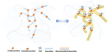
For the first time, a biohybrid nanofibrous wound dressing is developed via green electrospinning of a blend solution of bovine serum albumin (BSA) (1 and 3 wt%) and polycaprolactone (PCL). In such a system, the components are miscible and interact through hydrogen bonding between the carbonyl group of PCL and the amine group of BSA, as verified by ATR-FTIR. As a result, the biohybrid nanofibers show a superior elastic modulus and elongation (300% and 58%, respectively) compared with the neat PCL nanofibers. The included protein induces a hydrophilicity effect to the PCL nanofibers, notably at the higher BSA content (3 wt%). In contrast to the neat nanofibers, the biohybrid ones are bioactive and encourage formation of biominerals (made of amorphous calcium carbonate) on the surface, after immersion in simulated body fluid (SBF). Based on the WST-8 cell viability tests, NIH3T3 fibroblast cells were seen to properly interact with the biohybrid mats and to proliferate in their proximity. SEM images show that the cells largely adhere onto such nanofibers even more than they do on the neat ones and adopt a flattened and stretched shape. In addition, the live/dead assay and phalloidin/DAPI staining assay confirm large cell viability and normal cell morphology on the biohybrid nanofiber mats after 4 days incubation. Taken together, BSA/PCL nanofibers are able to offer optimum mechanical properties (elasticity) as well as mineralization which can potentially stimulate the wound healing process, and can be considered a suitable candidate for wound dressing applications.
中文翻译:
包含牛血清白蛋白作为伤口敷料生物活性部分的仿生生物杂化纳米纤维
第一次,通过绿色电纺牛血清白蛋白(BSA)(1和3 wt%)和聚己内酯(PCL)的混合溶液,开发了一种生物混合型纳米纤维伤口敷料。在这种系统中,各组分可混溶并通过PCL的羰基和BSA的胺基之间的氢键相互作用,这已通过ATR-FTIR验证。结果,与纯净的PCL纳米纤维相比,生物杂化纳米纤维显示出优异的弹性模量和伸长率(分别为300%和58%)。包含的蛋白质对PCL纳米纤维具有亲水作用,尤其是在较高的BSA含量(3 wt%)下。与纯净的纳米纤维相比,生物混合纤维具有生物活性,并且在浸入模拟体液(SBF)后会在表面形成生物矿物(由无定形碳酸钙制成)。根据WST-8细胞活力测试,NIH3T3成纤维细胞可与生物杂交垫正确相互作用并在其附近增殖。扫描电镜图像显示,细胞在纳米纤维上的粘附力甚至大于在纯净纤维上的粘附力,并呈现出扁平化和拉伸的形状。此外,在孵育4天后,活/死分析和鬼笔环肽/ DAPI染色分析证实了生物杂化纳米纤维垫上的大细胞活力和正常细胞形态。综上所述,BSA / PCL纳米纤维能够提供最佳的机械性能(弹性)以及矿化作用,从而有可能刺激伤口的愈合过程,并被认为是伤口敷料应用的合适候选者。观察到NIH3T3成纤维细胞与生物杂交垫正确相互作用并在其附近增殖。扫描电镜图像显示,细胞在纳米纤维上的粘附力甚至大于在纯净纤维上的粘附力,并呈现出扁平化和拉伸的形状。此外,在孵育4天后,活/死分析和鬼笔环肽/ DAPI染色分析证实了生物杂化纳米纤维垫上的大细胞活力和正常细胞形态。综上所述,BSA / PCL纳米纤维能够提供最佳的机械性能(弹性)以及矿化作用,从而有可能刺激伤口的愈合过程,并被认为是伤口敷料应用的合适候选者。观察到NIH3T3成纤维细胞与生物杂交垫正确相互作用并在其附近增殖。扫描电镜图像显示,细胞在纳米纤维上的粘附力甚至大于在纯净纤维上的粘附力,并呈现出扁平化和拉伸的形状。此外,在孵育4天后,活/死分析和鬼笔环肽/ DAPI染色分析证实了生物杂化纳米纤维垫上的大细胞活力和正常细胞形态。综上所述,BSA / PCL纳米纤维能够提供最佳的机械性能(弹性)以及矿化作用,从而有可能刺激伤口的愈合过程,并被认为是伤口敷料应用的合适候选者。扫描电镜图像显示,细胞在纳米纤维上的粘附力甚至大于在纯净纤维上的粘附力,并呈现出扁平化和拉伸的形状。此外,在孵育4天后,活/死分析和鬼笔环肽/ DAPI染色分析证实了生物杂化纳米纤维垫上的大细胞活力和正常细胞形态。综上所述,BSA / PCL纳米纤维能够提供最佳的机械性能(弹性)以及矿化作用,从而有可能刺激伤口的愈合过程,并被认为是伤口敷料应用的合适候选者。扫描电镜图像显示,细胞在纳米纤维上的粘附力甚至大于在纯净纤维上的粘附力,并呈现出扁平化和拉伸的形状。此外,在孵育4天后,活/死分析和鬼笔环肽/ DAPI染色分析证实了生物杂化纳米纤维垫上的大细胞活力和正常细胞形态。综上所述,BSA / PCL纳米纤维能够提供最佳的机械性能(弹性)以及矿化作用,从而有可能刺激伤口的愈合过程,并被认为是伤口敷料应用的合适候选者。


 京公网安备 11010802027423号