当前位置:
X-MOL 学术
›
Int. J. Dev. Neurosci.
›
论文详情
Our official English website, www.x-mol.net, welcomes your
feedback! (Note: you will need to create a separate account there.)
Signaling pathways modulated by monocular enucleation in the superior colliculus of juvenile rats
International Journal of Developmental Neuroscience ( IF 1.7 ) Pub Date : 2021-02-05 , DOI: 10.1002/jdn.10095 Juliana Ferreira Vasques 1, 2 , Renata Guedes de Jesus Gonçalves 1, 2 , Ana Lucia Tavares Gomes 1 , Paula Campello-Costa 1 , Claudio Alberto Serfaty 1 , Adriana da Cunha Faria-Melibeu 1
International Journal of Developmental Neuroscience ( IF 1.7 ) Pub Date : 2021-02-05 , DOI: 10.1002/jdn.10095 Juliana Ferreira Vasques 1, 2 , Renata Guedes de Jesus Gonçalves 1, 2 , Ana Lucia Tavares Gomes 1 , Paula Campello-Costa 1 , Claudio Alberto Serfaty 1 , Adriana da Cunha Faria-Melibeu 1
Affiliation
|
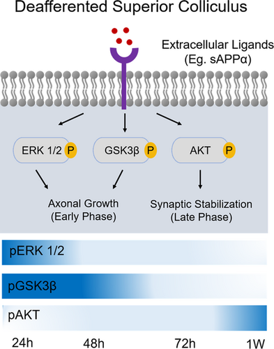
Monocular eye enucleation (ME) is a classical paradigm to induce neural plasticity in retinal ganglion cells (RGCs) axons from the intact eye, especially when performed within the critical period of visual system development. However, the precise mechanisms underlying the axonal sprouting and synaptogenesis seen in this model remain poorly understood. In the present work, we investigated the temporal alterations in phosphorylation of three kinases related to axonal growth and synaptogenesis—GSK3β (an important repressor of axonal outgrowth), AKT, and ERK—in superior colliculus of rats submitted to ME during early postnatal development. Western blotting analysis showed an increase in pGSK3β, the inactive form of this enzyme, 24 and 48 hr after ME. Accordingly, an increase in pERK levels was detected 24 hr after ME, indicating that phosphorylation of these enzymes might be related to axonal reorganization induced by ME. Interestingly, AKT phosphorylation was increased just 1 week after ME, suggesting it may be involved in the stabilization of newly formed synapses, rising from the axonal reorganization of remaining eye. A better understanding of how signaling pathways are modulated in a model of intense axonal sprouting can highlight possible therapeutic targets in RGCs injuries in adult individuals, where axonal regrowth is nearly absent.
中文翻译:

幼鼠上丘单眼摘除术调控的信号通路
单眼眼球摘除术 (ME) 是一种经典范式,可诱导来自完整眼睛的视网膜神经节细胞 (RGC) 轴突的神经可塑性,尤其是在视觉系统发育的关键时期进行时。然而,在该模型中看到的轴突萌发和突触发生的确切机制仍然知之甚少。在目前的工作中,我们研究了与轴突生长和突触发生相关的三种激酶——GSK3β(轴突生长的重要抑制因子)、AKT 和 ERK——在出生后早期发育期间接受 ME 的大鼠的上丘磷酸化的时间变化。蛋白质印迹分析显示,在 ME 后 24 和 48 小时,pGSK3β(该酶的非活性形式)增加。因此,在 ME 后 24 小时检测到 pERK 水平增加,表明这些酶的磷酸化可能与 ME 诱导的轴突重组有关。有趣的是,在 ME 后仅 1 周 AKT 磷酸化就增加了,这表明它可能参与稳定新形成的突触,从剩余眼睛的轴突重组中上升。更好地了解如何在强烈的轴突萌芽模型中调节信号通路可以突出成年个体 RGC 损伤的可能治疗目标,其中轴突再生几乎不存在。
更新日期:2021-02-05
中文翻译:
幼鼠上丘单眼摘除术调控的信号通路
单眼眼球摘除术 (ME) 是一种经典范式,可诱导来自完整眼睛的视网膜神经节细胞 (RGC) 轴突的神经可塑性,尤其是在视觉系统发育的关键时期进行时。然而,在该模型中看到的轴突萌发和突触发生的确切机制仍然知之甚少。在目前的工作中,我们研究了与轴突生长和突触发生相关的三种激酶——GSK3β(轴突生长的重要抑制因子)、AKT 和 ERK——在出生后早期发育期间接受 ME 的大鼠的上丘磷酸化的时间变化。蛋白质印迹分析显示,在 ME 后 24 和 48 小时,pGSK3β(该酶的非活性形式)增加。因此,在 ME 后 24 小时检测到 pERK 水平增加,表明这些酶的磷酸化可能与 ME 诱导的轴突重组有关。有趣的是,在 ME 后仅 1 周 AKT 磷酸化就增加了,这表明它可能参与稳定新形成的突触,从剩余眼睛的轴突重组中上升。更好地了解如何在强烈的轴突萌芽模型中调节信号通路可以突出成年个体 RGC 损伤的可能治疗目标,其中轴突再生几乎不存在。


 京公网安备 11010802027423号