当前位置:
X-MOL 学术
›
Microsc. Res. Tech.
›
论文详情
Our official English website, www.x-mol.net, welcomes your feedback! (Note: you will need to create a separate account there.)
Evaluation of surface roughness after root resection: An optical profilometer study
Microscopy Research and Technique ( IF 2.5 ) Pub Date : 2021-01-25 , DOI: 10.1002/jemt.23714 Ömer Ekici 1 , Kubilay Aslantaş 2 , Özgür Kanık 3 , Ali Keleş 4
Microscopy Research and Technique ( IF 2.5 ) Pub Date : 2021-01-25 , DOI: 10.1002/jemt.23714 Ömer Ekici 1 , Kubilay Aslantaş 2 , Özgür Kanık 3 , Ali Keleş 4
Affiliation
|
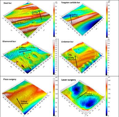
The aim of this study was to evaluate the roughness of the apical surface after apical resection performed by six different methods with an optical profilometer. Sixty human single root premolar teeth were used in this in vitro study. After root canal preparation, root canals were filled with gutta‐percha and AH Plus root canal sealers by lateral condensation technique. The teeth were randomly divided into six groups according to the apical resection method: steel fissure bur, tungsten carbide fissure bur, Lindeman bur, diamond fissure bur, laser, and ultrasonic surgical piezo with a diamond tip. The root ends were resected 3 mm away from the root apex and at a 90° angle. The time required for apicectomy was recorded for each group. After apical resection, the root surfaces were analyzed by an optical profilometer. The Kruskal–Wallis method was used to analyze the differences between groups. The significance level was set at 5%. The roughest surfaces were obtained by laser (25.54 ± 9.01 μm) and Lindeman bur (17.35 ± 6.03 μm), respectively. The longest mean resection times were recorded in piezosurgery and laser surgery (57 ± 14.39 s and 50.9 ± 16.86 s), respectively. Although the diamond‐tipped piezo surgical cutting time is long, it has the best results in terms of surface roughness (5.50 ± 1.73 μm). The optical profilometer is a more convenient tool for evaluating the surface after apical surgery, as it provides an opportunity to evaluate objectively with both visual and numerical data.
中文翻译:

根部切除后表面粗糙度的评估:光学轮廓仪研究
本研究的目的是使用光学轮廓仪评估通过六种不同方法进行的根尖切除后根尖表面的粗糙度。在这项体外研究中使用了 60 颗人类单根前磨牙。根管预备后,用牙胶和 AH Plus 根管封闭剂通过侧向冷凝技术填充根管。根据根尖切除方法将牙齿随机分为六组:钢裂隙车针、碳化钨裂隙车针、Lindeman 车针、金刚石裂隙车针、激光和带有金刚石尖端的超声外科压电。在距根尖 3 mm 处以 90° 角切除根端。记录每组根尖切除术所需的时间。根尖切除后,通过光学轮廓仪分析根表面。Kruskal-Wallis 方法用于分析组间差异。显着性水平设定为 5%。最粗糙的表面分别通过激光 (25.54 ± 9.01 μm) 和 Lindeman bur (17.35 ± 6.03 μm) 获得。在压电手术和激光手术中记录了最长的平均切除时间(分别为 57 ± 14.39 秒和 50.9 ± 16.86 秒)。虽然金刚石尖头压电手术切割时间较长,但在表面粗糙度(5.50 ± 1.73 μm)方面效果最好。光学轮廓仪是一种更方便的工具,用于评估根尖手术后的表面,因为它提供了使用视觉和数字数据进行客观评估的机会。35±6.03 微米),分别。在压电手术和激光手术中记录了最长的平均切除时间(分别为 57 ± 14.39 秒和 50.9 ± 16.86 秒)。虽然金刚石尖头压电手术切割时间较长,但在表面粗糙度(5.50 ± 1.73 μm)方面效果最好。光学轮廓仪是一种更方便的工具,用于评估根尖手术后的表面,因为它提供了使用视觉和数字数据进行客观评估的机会。35±6.03 微米),分别。在压电手术和激光手术中记录了最长的平均切除时间(分别为 57 ± 14.39 秒和 50.9 ± 16.86 秒)。虽然金刚石尖头压电手术切割时间较长,但在表面粗糙度(5.50 ± 1.73 μm)方面效果最好。光学轮廓仪是一种更方便的工具,用于评估根尖手术后的表面,因为它提供了使用视觉和数字数据进行客观评估的机会。
更新日期:2021-03-27
中文翻译:
根部切除后表面粗糙度的评估:光学轮廓仪研究
本研究的目的是使用光学轮廓仪评估通过六种不同方法进行的根尖切除后根尖表面的粗糙度。在这项体外研究中使用了 60 颗人类单根前磨牙。根管预备后,用牙胶和 AH Plus 根管封闭剂通过侧向冷凝技术填充根管。根据根尖切除方法将牙齿随机分为六组:钢裂隙车针、碳化钨裂隙车针、Lindeman 车针、金刚石裂隙车针、激光和带有金刚石尖端的超声外科压电。在距根尖 3 mm 处以 90° 角切除根端。记录每组根尖切除术所需的时间。根尖切除后,通过光学轮廓仪分析根表面。Kruskal-Wallis 方法用于分析组间差异。显着性水平设定为 5%。最粗糙的表面分别通过激光 (25.54 ± 9.01 μm) 和 Lindeman bur (17.35 ± 6.03 μm) 获得。在压电手术和激光手术中记录了最长的平均切除时间(分别为 57 ± 14.39 秒和 50.9 ± 16.86 秒)。虽然金刚石尖头压电手术切割时间较长,但在表面粗糙度(5.50 ± 1.73 μm)方面效果最好。光学轮廓仪是一种更方便的工具,用于评估根尖手术后的表面,因为它提供了使用视觉和数字数据进行客观评估的机会。35±6.03 微米),分别。在压电手术和激光手术中记录了最长的平均切除时间(分别为 57 ± 14.39 秒和 50.9 ± 16.86 秒)。虽然金刚石尖头压电手术切割时间较长,但在表面粗糙度(5.50 ± 1.73 μm)方面效果最好。光学轮廓仪是一种更方便的工具,用于评估根尖手术后的表面,因为它提供了使用视觉和数字数据进行客观评估的机会。35±6.03 微米),分别。在压电手术和激光手术中记录了最长的平均切除时间(分别为 57 ± 14.39 秒和 50.9 ± 16.86 秒)。虽然金刚石尖头压电手术切割时间较长,但在表面粗糙度(5.50 ± 1.73 μm)方面效果最好。光学轮廓仪是一种更方便的工具,用于评估根尖手术后的表面,因为它提供了使用视觉和数字数据进行客观评估的机会。


 京公网安备 11010802027423号