Cell Biology and Toxicology ( IF 5.3 ) Pub Date : 2021-01-21 , DOI: 10.1007/s10565-020-09575-9 Minghao Yan 1, 2 , Shen Gu 1, 2, 3 , Chun Pan 1, 2 , Yabing Chen 1, 2 , Xiaodong Han 1, 2
|
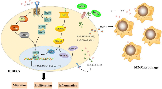
Microcystin-leucine-arginine (MC-LR) was produced by toxic cyanobacteria, which has been shown to have potent hepatotoxicity. Our previous study has proved that MC-LR were able to promote intrahepatic biliary epithelial cell excessive proliferation. However, the underlying mechanism is not yet entirely clarified. Herein, mice were fed with different concentrations (1, 7.5, 15, or 30 μg/L) of MC-LR by drinking water for 6 months. As the concentration of MC-LR increased, a growing number of macrophages were evaluated in the portal area of the mouse liver. Next, we built a co-culture system to explore the interaction between macrophages (THP-1 cells) and human intrahepatic biliary epithelial cells (HiBECs) in the presence of MC-LR. Under the exposure of MC-LR, HiBECs secreted a large amount of inflammatory factors (IL-6, IL-8, IL-1β, COX-2, XCL-1) and chemokine (MCP-1), which produced a huge chemotactic effect on THP-1 cells and induced elevation of the surface M2-subtype biomarkers (IL-10, CD163, CCL22, and Arg-1). In turn, high content of IL-6 in the medium activated JAK2/STAT3, MEK/ERK, and PI3K/AKT pathways in HiBECs, inducing HiBEC abnormal proliferation and migration. Together, these results suggested that MC-LR-mediated interaction between HiBECs and macrophages induced the M2-type polarization of macrophages, and activated IL-6/JAK2/STAT3, MEK/ERK, and PI3K/AKT pathways in HiBECs, further enhanced cell proliferation, improved cell migration, and hindered cell apoptosis by activating p-STAT3.
Graphical abstract
中文翻译:
MC-LR诱导的M2巨噬细胞与胆管上皮细胞相互作用通过调节STAT3促进胆管上皮细胞增殖和迁移
微囊藻毒素-亮氨酸-精氨酸 (MC-LR) 由有毒蓝藻产生,已被证明具有强肝毒性。我们前期的研究证明MC-LR能够促进肝内胆管上皮细胞过度增殖。然而,潜在的机制尚未完全阐明。在此,通过饮水给小鼠喂食不同浓度(1、7.5、15 或 30 μg/L)的 MC-LR 6 个月。随着 MC-LR 浓度的增加,越来越多的巨噬细胞在小鼠肝脏的门静脉区域被评估。接下来,我们建立了一个共培养系统来探索巨噬细胞(THP-1 细胞)和人肝内胆管上皮细胞(HiBECs)在 MC-LR 存在下的相互作用。在 MC-LR 的作用下,HiBECs 分泌大量炎症因子(IL-6、IL-8、IL-1β、COX-2、XCL-1) 和趋化因子 (MCP-1),它们对 THP-1 细胞产生巨大的趋化作用并诱导表面 M2 亚型生物标志物(IL-10、CD163、CCL22 和 Arg-1)升高。反过来,培养基中高含量的 IL-6 激活 HiBECs 中的 JAK2/STAT3、MEK/ERK 和 PI3K/AKT 通路,诱导 HiBEC 异常增殖和迁移。总之,这些结果表明 MC-LR 介导的 HiBECs 和巨噬细胞之间的相互作用诱导巨噬细胞的 M2 型极化,并激活 HiBECs 中的 IL-6/JAK2/STAT3、MEK/ERK 和 PI3K/AKT 通路,进一步增强细胞增殖,改善细胞迁移,并通过激活 p-STAT3 阻碍细胞凋亡。培养基中高含量的 IL-6 激活 HiBECs 中的 JAK2/STAT3、MEK/ERK 和 PI3K/AKT 通路,诱导 HiBEC 异常增殖和迁移。总之,这些结果表明 MC-LR 介导的 HiBECs 和巨噬细胞之间的相互作用诱导巨噬细胞的 M2 型极化,并激活 HiBECs 中的 IL-6/JAK2/STAT3、MEK/ERK 和 PI3K/AKT 通路,进一步增强细胞增殖,改善细胞迁移,并通过激活 p-STAT3 阻碍细胞凋亡。培养基中高含量的 IL-6 激活 HiBECs 中的 JAK2/STAT3、MEK/ERK 和 PI3K/AKT 通路,诱导 HiBEC 异常增殖和迁移。总之,这些结果表明 MC-LR 介导的 HiBECs 和巨噬细胞之间的相互作用诱导巨噬细胞的 M2 型极化,并激活 HiBECs 中的 IL-6/JAK2/STAT3、MEK/ERK 和 PI3K/AKT 通路,进一步增强细胞增殖,改善细胞迁移,并通过激活 p-STAT3 阻碍细胞凋亡。


 京公网安备 11010802027423号