当前位置:
X-MOL 学术
›
J. Mater. Chem. B
›
论文详情
Our official English website, www.x-mol.net, welcomes your
feedback! (Note: you will need to create a separate account there.)
Gold nanoparticles enhance fluorescence signals by flow cytometry at low antibody concentrations
Journal of Materials Chemistry B ( IF 6.1 ) Pub Date : 2020-12-30 , DOI: 10.1039/d0tb02309d Daniela S. Reis 1, 2, 3 , Vivian L. de Oliveira 3, 4, 5 , Misael L. Silva 3, 6, 7, 8 , Roberto M. Paniago 2, 3, 9, 10 , Luiz O. Ladeira 2, 3, 9, 10 , Lidia M. Andrade 2, 3, 9, 10
Journal of Materials Chemistry B ( IF 6.1 ) Pub Date : 2020-12-30 , DOI: 10.1039/d0tb02309d Daniela S. Reis 1, 2, 3 , Vivian L. de Oliveira 3, 4, 5 , Misael L. Silva 3, 6, 7, 8 , Roberto M. Paniago 2, 3, 9, 10 , Luiz O. Ladeira 2, 3, 9, 10 , Lidia M. Andrade 2, 3, 9, 10
Affiliation
|
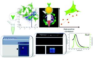
Flow cytometry is a universally applied technique in many biological and clinical assays to evaluate cells, bacteria, parasites, and particles at a micrometre scale. More advanced flow cytometers can detect small molecules down to the nanometre scale that may identify intracellular nanostructures. Advancements in the field of nanobiotechnology have led to techniques that allow the study of cellular behaviour after exposure to nanomaterials, particularly, metal nanoparticles. The optical properties of gold nanoparticles regarding surface plasmon resonance (SPR) are established to increase the fluorescence quantum yields of several dyes working as optical antennas, enabling the enhancement of light emission in fluorescent emitters. In this work we constructed a nanoprobe using gold nanoparticles coated with primary antibody Cetuximab. Then, we investigated whether this nanoprobe labelled with secondary fluorescent antibody Alexa Fluor 488, at low concentrations, could promote fluorescent signal enhancement, associated with SPR, and detected by the flow cytometry technique. Our results showed an enhanced fluorescent signal likely due to the proximity between the extinction coefficient of gold nanoparticles and the emission peak of Alexa Fluor 488, at exceptionally low concentrations, occurring within a high level of specificity. Moreover, the nanoprobe did not alter the cellular viability suggesting gold nanoparticles as a feasible approach for cell labelling using low concentrations of secondary antibodies for routine flow cytometry applications.
中文翻译:

金纳米颗粒通过低抗体浓度的流式细胞仪增强荧光信号
流式细胞术是在许多生物学和临床测定中普遍应用的技术,用于以微米为单位评估细胞,细菌,寄生虫和颗粒。更先进的流式细胞仪可以检测到可以识别细胞内纳米结构的纳米级小分子。纳米生物技术领域的进步导致了可以研究暴露于纳米材料(尤其是金属纳米颗粒)后的细胞行为的技术。建立有关表面等离子体共振(SPR)的金纳米颗粒的光学特性,以提高用作光学天线的几种染料的荧光量子产率,从而增强荧光发射器中的光发射。在这项工作中,我们使用涂有一抗西妥昔单抗的金纳米颗粒构建了纳米探针。然后,我们研究了用低浓度的次级荧光抗体Alexa Fluor 488标记的这种纳米探针是否可以促进与SPR相关的荧光信号增强,并通过流式细胞仪技术进行检测。我们的结果表明,荧光信号增强的原因可能是金纳米颗粒的消光系数与Alexa Fluor 488的发射峰之间的接近度,在极低的浓度下,其特异性很高。此外,纳米探针并没有改变细胞的生存能力,这表明金纳米颗粒是用于常规流式细胞术应用中低浓度二抗的细胞标记的可行方法。可以促进荧光信号增强,与SPR相关,并通过流式细胞仪技术检测。我们的结果表明,荧光信号增强的原因可能是金纳米颗粒的消光系数与Alexa Fluor 488的发射峰之间的接近度,在极低的浓度下,其特异性很高。此外,纳米探针并没有改变细胞的生存能力,这表明金纳米颗粒是用于常规流式细胞术应用中低浓度二抗的细胞标记的可行方法。可以促进荧光信号增强,与SPR相关,并通过流式细胞仪技术检测。我们的结果表明,荧光信号增强的原因可能是金纳米颗粒的消光系数与Alexa Fluor 488的发射峰之间的接近度,在极低的浓度下,其特异性很高。此外,纳米探针并没有改变细胞的生存能力,这表明金纳米颗粒是用于常规流式细胞术应用中低浓度二抗的细胞标记的可行方法。在极低的浓度下,以高度的特异性发生。此外,纳米探针并没有改变细胞的生存能力,这表明金纳米颗粒是用于常规流式细胞术应用中低浓度二抗的细胞标记的可行方法。浓度极低,发生在高特异性范围内。此外,纳米探针并没有改变细胞的生存能力,这表明金纳米颗粒是用于常规流式细胞术应用的低浓度第二抗体进行细胞标记的可行方法。
更新日期:2021-01-19
中文翻译:
金纳米颗粒通过低抗体浓度的流式细胞仪增强荧光信号
流式细胞术是在许多生物学和临床测定中普遍应用的技术,用于以微米为单位评估细胞,细菌,寄生虫和颗粒。更先进的流式细胞仪可以检测到可以识别细胞内纳米结构的纳米级小分子。纳米生物技术领域的进步导致了可以研究暴露于纳米材料(尤其是金属纳米颗粒)后的细胞行为的技术。建立有关表面等离子体共振(SPR)的金纳米颗粒的光学特性,以提高用作光学天线的几种染料的荧光量子产率,从而增强荧光发射器中的光发射。在这项工作中,我们使用涂有一抗西妥昔单抗的金纳米颗粒构建了纳米探针。然后,我们研究了用低浓度的次级荧光抗体Alexa Fluor 488标记的这种纳米探针是否可以促进与SPR相关的荧光信号增强,并通过流式细胞仪技术进行检测。我们的结果表明,荧光信号增强的原因可能是金纳米颗粒的消光系数与Alexa Fluor 488的发射峰之间的接近度,在极低的浓度下,其特异性很高。此外,纳米探针并没有改变细胞的生存能力,这表明金纳米颗粒是用于常规流式细胞术应用中低浓度二抗的细胞标记的可行方法。可以促进荧光信号增强,与SPR相关,并通过流式细胞仪技术检测。我们的结果表明,荧光信号增强的原因可能是金纳米颗粒的消光系数与Alexa Fluor 488的发射峰之间的接近度,在极低的浓度下,其特异性很高。此外,纳米探针并没有改变细胞的生存能力,这表明金纳米颗粒是用于常规流式细胞术应用中低浓度二抗的细胞标记的可行方法。可以促进荧光信号增强,与SPR相关,并通过流式细胞仪技术检测。我们的结果表明,荧光信号增强的原因可能是金纳米颗粒的消光系数与Alexa Fluor 488的发射峰之间的接近度,在极低的浓度下,其特异性很高。此外,纳米探针并没有改变细胞的生存能力,这表明金纳米颗粒是用于常规流式细胞术应用中低浓度二抗的细胞标记的可行方法。在极低的浓度下,以高度的特异性发生。此外,纳米探针并没有改变细胞的生存能力,这表明金纳米颗粒是用于常规流式细胞术应用中低浓度二抗的细胞标记的可行方法。浓度极低,发生在高特异性范围内。此外,纳米探针并没有改变细胞的生存能力,这表明金纳米颗粒是用于常规流式细胞术应用的低浓度第二抗体进行细胞标记的可行方法。


 京公网安备 11010802027423号