Computer Methods and Programs in Biomedicine ( IF 4.9 ) Pub Date : 2021-01-19 , DOI: 10.1016/j.cmpb.2021.105946 Sedighe Firuzinia , Seyed Mahmoodreza Afzali , Fatemeh Ghasemian , Seyed Abolghasem Mirroshandel
|
Background and objective
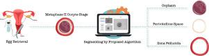
The morphology of the human metaphase II (MII) oocyte is an essential indicator of the embryo's potential for developing into a healthy baby in the Intra-Cytoplasmic Sperm Injection (ICSI) process. In this case, characteristics such as oocyte and ooplasm area, zona pellucida (ZP) thickness, and perivitelline space (PVS) width are also linked to the embryo's implantation potential. Moreover, oocyte segmentation methods may be of particular interest in those countries' restrictive IVF legislation.
Methods
While the manual examination is impractically time-consuming and subjective, this paper concentrates efforts on designing an automated deep learning framework to take on the challenging task of segmentation in low-resolution microscopic images of MII oocytes. In particular, we have developed a deep learning network based on an improved U-Net model using our presented unique collection of human MII oocyte images (a new challenging dataset contains 1,009 images accompanied by manually labeled pixel-accurate ground truths). High-quality ground truth (GT) preparation is a labor-intensive task. However, we put considerable effort into assessing how different types of GT annotations (binary and multiclass) impact segmentation performance.
Results
Experimental results on 250 MII oocyte test images demonstrate that the proposed multiclass segmentation algorithm is able to segment complex and irregular ooplasm, ZP, and PVS structures more accurately than its two-class version. Furthermore, the proposed architecture outperforms two other state-of-the-art deep learning models, U-Net and ENet, for the MII oocyte segmentation task.
Conclusions
The findings of this study provide a fascinating insight into the automatic and accurate segmentation of human MII oocytes.
中文翻译:
基于稳健的深度学习的多类分割方法,用于分析人类中期II卵母细胞图像
背景和目标
人类中期II(MII)卵母细胞的形态是胚胎在细胞质内精子注射(ICSI)过程中发展为健康婴儿的潜力的重要指标。在这种情况下,诸如卵母细胞和卵母细胞的面积,透明带(Zona pellucida,ZP)的厚度和玻璃周间隙(PVS)的宽度等特征也与胚胎的植入潜力有关。此外,卵母细胞分割方法可能在这些国家的限制性IVF法规中特别有用。
方法
尽管手动检查是不切实际的耗时且主观的,但本文集中精力设计了一个自动化的深度学习框架,以应对MII卵母细胞低分辨率显微图像中具有挑战性的分割任务。尤其是,我们已经使用我们提出的人类MII卵母细胞图像的独特集合(基于新的具有挑战性的数据集,包含1,009张图像以及手动标记的像素准确的地面真相),基于改进的U-Net模型开发了深度学习网络。高质量的地面真相(GT)准备工作是一项劳动密集型任务。但是,我们投入了大量精力来评估不同类型的GT注释(二进制和多类)如何影响细分效果。
结果
在250个MII卵母细胞测试图像上的实验结果表明,所提出的多类分割算法比其两类版本能够更准确地分割复杂和不规则的卵质,ZP和PVS结构。此外,针对MII卵母细胞分割任务,拟议的架构优于其他两个最先进的深度学习模型U-Net和ENet。
结论
这项研究的发现为人类MII卵母细胞的自动和准确分割提供了令人着迷的见解。


 京公网安备 11010802027423号