当前位置:
X-MOL 学术
›
Anal. Methods
›
论文详情
Our official English website, www.x-mol.net, welcomes your
feedback! (Note: you will need to create a separate account there.)
Quantitative proteomics of epigenetic histone modifications in MCF-7 cells under estradiol stimulation
Analytical Methods ( IF 2.7 ) Pub Date : 2020-12-18 , DOI: 10.1039/d0ay02146f Yechen Hu 1, 2, 3, 4, 5 , Hao Jiang 1, 2, 3, 4, 5 , Baofeng Zhao 1, 2, 3, 4, 5 , Kaiguang Yang 1, 2, 3, 4, 5 , Zhen Liang 1, 2, 3, 4, 5 , Lihua Zhang 1, 2, 3, 4, 5 , Yukui Zhang 1, 2, 3, 4, 5
Analytical Methods ( IF 2.7 ) Pub Date : 2020-12-18 , DOI: 10.1039/d0ay02146f Yechen Hu 1, 2, 3, 4, 5 , Hao Jiang 1, 2, 3, 4, 5 , Baofeng Zhao 1, 2, 3, 4, 5 , Kaiguang Yang 1, 2, 3, 4, 5 , Zhen Liang 1, 2, 3, 4, 5 , Lihua Zhang 1, 2, 3, 4, 5 , Yukui Zhang 1, 2, 3, 4, 5
Affiliation
|
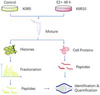
Estrogen exposure has already been considered to be associated with tumorigenesis and breast cancer progression. To study the epigenetic regulation mechanism in MCF-7 cells under estrogen exposure, which normally results in cell proliferation and malignancy, a stable isotope labeling of amino acid (SILAC) based quantitative proteomics strategy was used to analyse histone post-translational modifications (PTMs) and protein differential expressions. In total, we have unambiguously identified 49 histone variants and quantified 42 of them, in which two differentially expressed proteins were found to be associated with breast cancers. Through the quantitative analysis of 470 histone peptides with a combination of different PTM types, including methylation (mono-, di-, and tri-), acetylation and phosphorylation, 150 of them were found to be differentially expressed. Through the biological analysis of the quantification results of both histone PTMs and proteins in MCF-7 cells, we found that (1) the histone variants H10 and H2AV have an effect on the adjustment of the nucleosome or chromatin structure and activate target genes; (2) after estrogen receptor (ER) activation by estrogen, the recruitment of histone acetyltransferase KAT7 might affect the acetylation at the N terminal of H4 (K5, K8 and K12) and also result in cross-talk between different acetylation sites; (3) different expression of histone deacetylase HDAC2 and its nucleo-cytoplasmic transportation process is important in the regulation of histone acetylation in MCF-7 cells under estrogen exposure.
中文翻译:

雌二醇刺激下MCF-7细胞表观遗传组蛋白修饰的定量蛋白质组学
雌激素暴露已被认为与肿瘤发生和乳腺癌进展有关。为了研究暴露于雌激素下通常会导致细胞增殖和恶性的MCF-7细胞的表观遗传调控机制,使用了基于氨基酸的稳定同位素标记(SILAC)的定量蛋白质组学策略来分析组蛋白翻译后修饰(PTM)和蛋白质差异表达。总共,我们明确地鉴定出49个组蛋白变体,并量化了其中的42个,其中发现两个差异表达的蛋白与乳腺癌有关。通过对470种组蛋白肽(包括甲基化(单,二和三),乙酰化和磷酸化)的不同PTM类型的组合进行定量分析,发现其中有150个差异表达。通过对MCF-7细胞中组蛋白PTM和蛋白质定量结果的生物学分析,我们发现(1)组蛋白变体H10和H2AV对核小体或染色质结构的调节和激活靶基因有影响;(2)在雌激素激活雌激素受体(ER)后,组蛋白乙酰转移酶KAT7的募集可能影响H4(K5,K8和K12)N末端的乙酰化,并导致不同乙酰化位点之间的串扰。(3)组蛋白脱乙酰基酶HDAC2的不同表达及其核质运输过程对调节雌激素暴露下MCF-7细胞组蛋白乙酰化具有重要意义。通过对MCF-7细胞中组蛋白PTM和蛋白质定量结果的生物学分析,我们发现(1)组蛋白变体H10和H2AV对核小体或染色质结构的调节和激活靶基因有影响;(2)在雌激素激活雌激素受体(ER)后,组蛋白乙酰转移酶KAT7的募集可能影响H4(K5,K8和K12)N末端的乙酰化,并导致不同乙酰化位点之间的串扰。(3)组蛋白脱乙酰基酶HDAC2的不同表达及其核质运输过程对调节雌激素暴露下MCF-7细胞组蛋白乙酰化具有重要意义。通过对MCF-7细胞中组蛋白PTM和蛋白质定量结果的生物学分析,我们发现(1)组蛋白变体H10和H2AV对核小体或染色质结构的调节和激活靶基因有影响;(2)在雌激素激活雌激素受体(ER)后,组蛋白乙酰转移酶KAT7的募集可能影响H4(K5,K8和K12)N末端的乙酰化,并导致不同乙酰化位点之间的串扰。(3)组蛋白脱乙酰基酶HDAC2的不同表达及其核质转运过程对暴露于雌激素的MCF-7细胞组蛋白乙酰化具有重要意义。我们发现(1)组蛋白变体H10和H2AV对核小体或染色质结构的调节有影响并激活靶基因;(2)在雌激素激活雌激素受体(ER)后,组蛋白乙酰转移酶KAT7的募集可能影响H4(K5,K8和K12)N末端的乙酰化,并导致不同乙酰化位点之间的串扰。(3)组蛋白脱乙酰基酶HDAC2的不同表达及其核质运输过程对调节雌激素暴露下MCF-7细胞组蛋白乙酰化具有重要意义。我们发现(1)组蛋白变体H10和H2AV对核小体或染色质结构的调节有影响并激活靶基因;(2)在雌激素激活雌激素受体(ER)后,组蛋白乙酰转移酶KAT7的募集可能影响H4(K5,K8和K12)N末端的乙酰化,并导致不同乙酰化位点之间的串扰。(3)组蛋白脱乙酰基酶HDAC2的不同表达及其核质运输过程对调节雌激素暴露下MCF-7细胞组蛋白乙酰化具有重要意义。K8和K12),还会导致不同的乙酰化位点之间发生串扰;(3)组蛋白脱乙酰基酶HDAC2的不同表达及其核质运输过程对调节雌激素暴露下MCF-7细胞组蛋白乙酰化具有重要意义。K8和K12),还会导致不同的乙酰化位点之间发生串扰;(3)组蛋白脱乙酰基酶HDAC2的不同表达及其核质运输过程对调节雌激素暴露下MCF-7细胞组蛋白乙酰化具有重要意义。
更新日期:2021-01-18
中文翻译:
雌二醇刺激下MCF-7细胞表观遗传组蛋白修饰的定量蛋白质组学
雌激素暴露已被认为与肿瘤发生和乳腺癌进展有关。为了研究暴露于雌激素下通常会导致细胞增殖和恶性的MCF-7细胞的表观遗传调控机制,使用了基于氨基酸的稳定同位素标记(SILAC)的定量蛋白质组学策略来分析组蛋白翻译后修饰(PTM)和蛋白质差异表达。总共,我们明确地鉴定出49个组蛋白变体,并量化了其中的42个,其中发现两个差异表达的蛋白与乳腺癌有关。通过对470种组蛋白肽(包括甲基化(单,二和三),乙酰化和磷酸化)的不同PTM类型的组合进行定量分析,发现其中有150个差异表达。通过对MCF-7细胞中组蛋白PTM和蛋白质定量结果的生物学分析,我们发现(1)组蛋白变体H10和H2AV对核小体或染色质结构的调节和激活靶基因有影响;(2)在雌激素激活雌激素受体(ER)后,组蛋白乙酰转移酶KAT7的募集可能影响H4(K5,K8和K12)N末端的乙酰化,并导致不同乙酰化位点之间的串扰。(3)组蛋白脱乙酰基酶HDAC2的不同表达及其核质运输过程对调节雌激素暴露下MCF-7细胞组蛋白乙酰化具有重要意义。通过对MCF-7细胞中组蛋白PTM和蛋白质定量结果的生物学分析,我们发现(1)组蛋白变体H10和H2AV对核小体或染色质结构的调节和激活靶基因有影响;(2)在雌激素激活雌激素受体(ER)后,组蛋白乙酰转移酶KAT7的募集可能影响H4(K5,K8和K12)N末端的乙酰化,并导致不同乙酰化位点之间的串扰。(3)组蛋白脱乙酰基酶HDAC2的不同表达及其核质运输过程对调节雌激素暴露下MCF-7细胞组蛋白乙酰化具有重要意义。通过对MCF-7细胞中组蛋白PTM和蛋白质定量结果的生物学分析,我们发现(1)组蛋白变体H10和H2AV对核小体或染色质结构的调节和激活靶基因有影响;(2)在雌激素激活雌激素受体(ER)后,组蛋白乙酰转移酶KAT7的募集可能影响H4(K5,K8和K12)N末端的乙酰化,并导致不同乙酰化位点之间的串扰。(3)组蛋白脱乙酰基酶HDAC2的不同表达及其核质转运过程对暴露于雌激素的MCF-7细胞组蛋白乙酰化具有重要意义。我们发现(1)组蛋白变体H10和H2AV对核小体或染色质结构的调节有影响并激活靶基因;(2)在雌激素激活雌激素受体(ER)后,组蛋白乙酰转移酶KAT7的募集可能影响H4(K5,K8和K12)N末端的乙酰化,并导致不同乙酰化位点之间的串扰。(3)组蛋白脱乙酰基酶HDAC2的不同表达及其核质运输过程对调节雌激素暴露下MCF-7细胞组蛋白乙酰化具有重要意义。我们发现(1)组蛋白变体H10和H2AV对核小体或染色质结构的调节有影响并激活靶基因;(2)在雌激素激活雌激素受体(ER)后,组蛋白乙酰转移酶KAT7的募集可能影响H4(K5,K8和K12)N末端的乙酰化,并导致不同乙酰化位点之间的串扰。(3)组蛋白脱乙酰基酶HDAC2的不同表达及其核质运输过程对调节雌激素暴露下MCF-7细胞组蛋白乙酰化具有重要意义。K8和K12),还会导致不同的乙酰化位点之间发生串扰;(3)组蛋白脱乙酰基酶HDAC2的不同表达及其核质运输过程对调节雌激素暴露下MCF-7细胞组蛋白乙酰化具有重要意义。K8和K12),还会导致不同的乙酰化位点之间发生串扰;(3)组蛋白脱乙酰基酶HDAC2的不同表达及其核质运输过程对调节雌激素暴露下MCF-7细胞组蛋白乙酰化具有重要意义。


 京公网安备 11010802027423号