当前位置:
X-MOL 学术
›
Biomacromolecules
›
论文详情
Our official English website, www.x-mol.net, welcomes your
feedback! (Note: you will need to create a separate account there.)
Surface-Functionalized Conducting Nanofibers for Electrically Stimulated Neural Cell Function
Biomacromolecules ( IF 5.5 ) Pub Date : 2021-01-15 , DOI: 10.1021/acs.biomac.0c01445 Rajiv Borah 1 , Ganesh C Ingavle 2 , Ashok Kumar 3 , Susan R Sandeman 4 , Sergey V Mikhalovsky 5, 6
Biomacromolecules ( IF 5.5 ) Pub Date : 2021-01-15 , DOI: 10.1021/acs.biomac.0c01445 Rajiv Borah 1 , Ganesh C Ingavle 2 , Ashok Kumar 3 , Susan R Sandeman 4 , Sergey V Mikhalovsky 5, 6
Affiliation

|
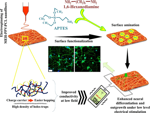
Strategies involving the inclusion of cell-instructive chemical and topographical cues to smart biomaterials in combination with a suitable physical stimulus may be beneficial to enhance nerve-regeneration rate. In this regard, we investigated the surface functionalization of poly[2-methoxy-5-(2-ethylhexyloxy)-1,4-phenylenevinylene] (MEH-PPV)-based electroconductive electrospun nanofibers coupled with externally applied electrical stimulus for accelerated neuronal growth potential. In addition, the voltage-dependent conductive mechanism of the nanofibers was studied in depth to interlink intrinsic conductive properties with electrically stimulated neuronal expressions. Surface functionalization was accomplished using 3-aminopropyltriethoxysilane (APTES) and 1,6-hexanediamine (HDA) as an alternative to costly biomolecule coating (e.g., collagen) for cell adhesion. The nanofibers were uniform, porous, electrically conductive, mechanically strong, and stable under physiological conditions. Surface amination boosted biocompatibility, 3T3 cell adhesion, and spreading, while the neuronal model rat PC12 cell line showed better differentiation on surface-functionalized mats compared to nonfunctionalized mats. When coupled with electrical stimulation (ES), these mats showed comparable or faster neurite formation and elongation than the collagen-coated mats with no-ES conditions. The findings indicate that surface amination in combination with ES may provide an improved strategy to faster nerve regeneration using MEH-PPV-based neural scaffolds.
中文翻译:

用于电刺激神经细胞功能的表面功能化导电纳米纤维
将细胞指导性化学和地形学线索包括在智能生物材料中并与适当的物理刺激相结合的策略可能对提高神经再生率有益。在这方面,我们研究了基于聚[2-甲氧基-5-(2-乙基己氧基)-1,4-亚苯基亚乙烯基](MEH-PPV)的导电电纺纳米纤维的表面功能化,并结合外部施加的电刺激来促进神经元生长潜在。此外,深入研究了纳米纤维的电压依赖性导电机理,以使固有的导电性能与电刺激的神经元表达相互关联。使用3-氨基丙基三乙氧基硅烷(APTES)和1,6-己二胺(HDA)作为昂贵的生物分子涂层(例如,胶原蛋白)用于细胞粘附。纳米纤维是均匀的,多孔的,导电的,机械强度的并且在生理条件下是稳定的。表面胺化可增强生物相容性,3T3细胞粘附和扩散,而神经元模型大鼠PC12细胞系与未功能化的垫相比在表面功能化的垫上表现出更好的分化。当与电刺激(ES)结合使用时,与无ES条件的胶原蛋白覆盖垫相比,这些垫显示出相当或更快的神经突形成和伸长。研究结果表明,表面胺化与ES结合使用基于MEH-PPV的神经支架可以提供更快的神经再生的改进策略。表面胺化可增强生物相容性,3T3细胞粘附和扩散,而神经元模型大鼠PC12细胞系与未功能化的垫相比在表面功能化的垫上表现出更好的分化。当与电刺激(ES)结合使用时,与无ES条件的胶原蛋白覆盖垫相比,这些垫显示出相当或更快的神经突形成和伸长。研究结果表明,表面胺化与ES结合使用基于MEH-PPV的神经支架可以提供更快的神经再生的改进策略。表面胺化可增强生物相容性,3T3细胞粘附和扩散,而神经元模型大鼠PC12细胞系与未功能化的垫相比在表面功能化的垫上表现出更好的分化。当与电刺激(ES)结合使用时,与无ES条件的胶原蛋白覆盖垫相比,这些垫显示出相当或更快的神经突形成和伸长。研究结果表明,表面胺化与ES结合使用基于MEH-PPV的神经支架可以提供更快的神经再生的改进策略。这些垫子在没有ES的条件下显示出与胶原蛋白涂覆的垫子相当或更快的神经突形成和伸长。研究结果表明,表面胺化与ES结合可以为使用基于MEH-PPV的神经支架提供更快的神经再生提供更好的策略。这些垫子在没有ES的条件下显示出与胶原蛋白涂覆的垫子相当或更快的神经突形成和伸长。研究结果表明,表面胺化与ES结合使用基于MEH-PPV的神经支架可以提供更快的神经再生的改进策略。
更新日期:2021-02-08
中文翻译:

用于电刺激神经细胞功能的表面功能化导电纳米纤维
将细胞指导性化学和地形学线索包括在智能生物材料中并与适当的物理刺激相结合的策略可能对提高神经再生率有益。在这方面,我们研究了基于聚[2-甲氧基-5-(2-乙基己氧基)-1,4-亚苯基亚乙烯基](MEH-PPV)的导电电纺纳米纤维的表面功能化,并结合外部施加的电刺激来促进神经元生长潜在。此外,深入研究了纳米纤维的电压依赖性导电机理,以使固有的导电性能与电刺激的神经元表达相互关联。使用3-氨基丙基三乙氧基硅烷(APTES)和1,6-己二胺(HDA)作为昂贵的生物分子涂层(例如,胶原蛋白)用于细胞粘附。纳米纤维是均匀的,多孔的,导电的,机械强度的并且在生理条件下是稳定的。表面胺化可增强生物相容性,3T3细胞粘附和扩散,而神经元模型大鼠PC12细胞系与未功能化的垫相比在表面功能化的垫上表现出更好的分化。当与电刺激(ES)结合使用时,与无ES条件的胶原蛋白覆盖垫相比,这些垫显示出相当或更快的神经突形成和伸长。研究结果表明,表面胺化与ES结合使用基于MEH-PPV的神经支架可以提供更快的神经再生的改进策略。表面胺化可增强生物相容性,3T3细胞粘附和扩散,而神经元模型大鼠PC12细胞系与未功能化的垫相比在表面功能化的垫上表现出更好的分化。当与电刺激(ES)结合使用时,与无ES条件的胶原蛋白覆盖垫相比,这些垫显示出相当或更快的神经突形成和伸长。研究结果表明,表面胺化与ES结合使用基于MEH-PPV的神经支架可以提供更快的神经再生的改进策略。表面胺化可增强生物相容性,3T3细胞粘附和扩散,而神经元模型大鼠PC12细胞系与未功能化的垫相比在表面功能化的垫上表现出更好的分化。当与电刺激(ES)结合使用时,与无ES条件的胶原蛋白覆盖垫相比,这些垫显示出相当或更快的神经突形成和伸长。研究结果表明,表面胺化与ES结合使用基于MEH-PPV的神经支架可以提供更快的神经再生的改进策略。这些垫子在没有ES的条件下显示出与胶原蛋白涂覆的垫子相当或更快的神经突形成和伸长。研究结果表明,表面胺化与ES结合可以为使用基于MEH-PPV的神经支架提供更快的神经再生提供更好的策略。这些垫子在没有ES的条件下显示出与胶原蛋白涂覆的垫子相当或更快的神经突形成和伸长。研究结果表明,表面胺化与ES结合使用基于MEH-PPV的神经支架可以提供更快的神经再生的改进策略。


 京公网安备 11010802027423号