当前位置:
X-MOL 学术
›
J. Synchrotron Radiat.
›
论文详情
Our official English website, www.x-mol.net, welcomes your
feedback! (Note: you will need to create a separate account there.)
Oxidation‐induced three‐dimensional morphological changes in Ni nanoparticles observed by coherent X‐ray diffraction imaging
Journal of Synchrotron Radiation ( IF 2.4 ) Pub Date : 2021-01-14 , DOI: 10.1107/s1600577520015945 Kangwoo Ahn , In Hwa Cho , Junhyung Kim , Su Yong Lee , Daeho Sung , Chulho Jung , Changyong Song , Hyon Chol Kang , Do Young Noh
Journal of Synchrotron Radiation ( IF 2.4 ) Pub Date : 2021-01-14 , DOI: 10.1107/s1600577520015945 Kangwoo Ahn , In Hwa Cho , Junhyung Kim , Su Yong Lee , Daeho Sung , Chulho Jung , Changyong Song , Hyon Chol Kang , Do Young Noh
|
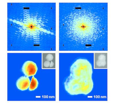
Three‐dimensional structures of Ni nanoparticles undergoing significant morphological changes on oxidation were observed non‐destructively using coherent X‐ray diffraction imaging. The Ni particles were oxidized into Ni1O1 while forming pores of various sizes internally. For each Ni nanoparticle, one large void was identified at a lower corner near the interface with the substrate. The porosity of the internal region of the agglomerated Ni oxide was about 38.4%. Regions of high NiO density were mostly observed at the outer crust of the oxide or at the boundary with the large voids. This research expands our understanding of general catalytic reactions with direct observation of oxidation‐induced nanoscale morphological changes.
中文翻译:

通过相干X射线衍射成像观察到的氧化镍纳米颗粒的三维形态变化
使用相干X射线衍射成像可以无损地观察到Ni纳米粒子在氧化过程中发生明显形态变化的三维结构。Ni粒子在内部形成各种大小的细孔的同时被氧化为Ni 1 O 1。对于每个Ni纳米颗粒,在与基底的界面附近的下角处发现了一个大的空隙。团聚的氧化镍的内部区域的孔隙率为约38.4%。高NiO密度的区域主要在氧化物的外壳或具有大空隙的边界处观察到。这项研究通过直接观察氧化引起的纳米级形态变化,扩展了我们对一般催化反应的理解。
更新日期:2021-03-04
中文翻译:
通过相干X射线衍射成像观察到的氧化镍纳米颗粒的三维形态变化
使用相干X射线衍射成像可以无损地观察到Ni纳米粒子在氧化过程中发生明显形态变化的三维结构。Ni粒子在内部形成各种大小的细孔的同时被氧化为Ni 1 O 1。对于每个Ni纳米颗粒,在与基底的界面附近的下角处发现了一个大的空隙。团聚的氧化镍的内部区域的孔隙率为约38.4%。高NiO密度的区域主要在氧化物的外壳或具有大空隙的边界处观察到。这项研究通过直接观察氧化引起的纳米级形态变化,扩展了我们对一般催化反应的理解。


 京公网安备 11010802027423号