NeuroImage: Clinical ( IF 3.4 ) Pub Date : 2021-01-14 , DOI: 10.1016/j.nicl.2021.102562 Sara Leddy 1 , Laura Serra 2 , Davide Esposito 3 , Camilla Vizzotto 3 , Giovanni Giulietti 2 , Gabriella Silvestri 4 , Antonio Petrucci 5 , Giovanni Meola 6 , Leonardo Lopiano 7 , Mara Cercignani 8 , Marco Bozzali 9
|
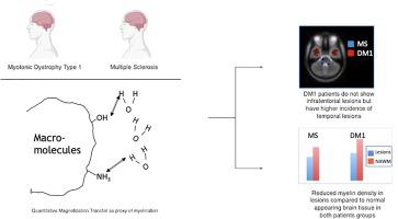
Myotonic Dystrophy type 1 (DM1) is an autosomal dominant condition caused by expansion of the CTG triplet repeats within the myotonic dystrophy protein of the kinase (DMPK) gene. The central nervous system is involved in the disease, with multiple symptoms including cognitive impairment. A typical feature of DM1 is the presence of widespread white matter (WM) lesions, whose total volume is associated with CTG triplet expansion. The aim of this study was to characterize the distribution and pathological substrate of these lesions as well as the normal appearing WM (NAWM) using quantitative magnetization transfer (qMT) MRI, and comparing data from DM1 patients with those from patients with multiple sclerosis (MS). Twenty-eight patients with DM1, 29 patients with relapsing-remitting MS, and 15 healthy controls had an MRI scan, including conventional and qMT imaging. The average pool size ratio (F), a proxy of myelination, was computed within lesions and NAWM for every participant. The lesion masks were warped into MNI space and lesion probability maps were obtained for each patient group. The lesion distribution, total lesion load and the tissue-specific mean F were compared between groups. The supratentorial distribution of lesions was similar in the 2 patient groups, although mean lesion volume was higher in MS than DM1. DM1 presented higher prevalence of anterior temporal lobe lesions, but none in the cerebellum and brainstem. Significantly reduced F values were found within DM1 lesions, suggesting a loss of myelin density. While F was reduced in the NAWM of MS patients, it did not differ between DM1 and controls. Our results provide further evidence for a need to compare histology and imaging using new MRI techniques in DM1 patients, in order to further our understanding of the underlying disease process contributing to WM disease.
中文翻译:
1型强直性肌营养不良症的病变分布和白质损伤的底物:与多发性硬化症的比较
1型强直性肌营养不良症(DM1)是常染色体显性疾病,由激酶(DMPK)基因的强直性肌营养不良蛋白内CTG三联体重复序列的扩增引起。该疾病涉及中枢神经系统,具有多种症状,包括认知障碍。DM1的典型特征是存在广泛的白质(WM)病变,其总体积与CTG三联体扩张相关。这项研究的目的是使用定量磁化转移(qMT)MRI表征这些病变以及正常出现的WM(NAWM)的分布和病理学基底,并比较DM1患者和多发性硬化症(MS)患者的数据)。进行MRI扫描的28例DM1患者,29例复发缓解型MS患者和15例健康对照者,包括常规和qMT成像。计算每个参与者在病变和NAWM内的平均池大小比率(F),这是髓鞘形成的代表。将病变罩翘曲到MNI空间中,并获得每个患者组的病变概率图。比较两组之间的病灶分布,总病灶负荷和组织特异性均值F。尽管MS中的平均病变体积比DM1高,但在两个患者组中病变的上皮分布相似。DM1表现出较高的前颞叶病变发生率,但在小脑和脑干中均没有。在DM1病变内发现F值显着降低,表明髓磷脂密度降低。尽管MS患者的NAWM中F降低,但DM1与对照组之间无差异。


 京公网安备 11010802027423号