当前位置:
X-MOL 学术
›
Hum. Brain Mapp.
›
论文详情
Our official English website, www.x-mol.net, welcomes your
feedback! (Note: you will need to create a separate account there.)
Evidence for interhemispheric imbalance in stroke patients as revealed by combining transcranial magnetic stimulation and electroencephalography
Human Brain Mapping ( IF 3.5 ) Pub Date : 2021-01-13 , DOI: 10.1002/hbm.25297 Elias Paolo Casula 1, 2 , Maria Concetta Pellicciari 1 , Sonia Bonnì 1 , Barbara Spanò 3 , Viviana Ponzo 1 , Ilenia Salsano 3 , Giovanni Giulietti 3 , Alex Martino Cinnera 1 , Michele Maiella 1 , Ilaria Borghi 1 , Lorenzo Rocchi 2 , Marco Bozzali 3, 4 , Fabrizio Sallustio 5 , Carlo Caltagirone 1 , Giacomo Koch 1, 5
Human Brain Mapping ( IF 3.5 ) Pub Date : 2021-01-13 , DOI: 10.1002/hbm.25297 Elias Paolo Casula 1, 2 , Maria Concetta Pellicciari 1 , Sonia Bonnì 1 , Barbara Spanò 3 , Viviana Ponzo 1 , Ilenia Salsano 3 , Giovanni Giulietti 3 , Alex Martino Cinnera 1 , Michele Maiella 1 , Ilaria Borghi 1 , Lorenzo Rocchi 2 , Marco Bozzali 3, 4 , Fabrizio Sallustio 5 , Carlo Caltagirone 1 , Giacomo Koch 1, 5
Affiliation
|
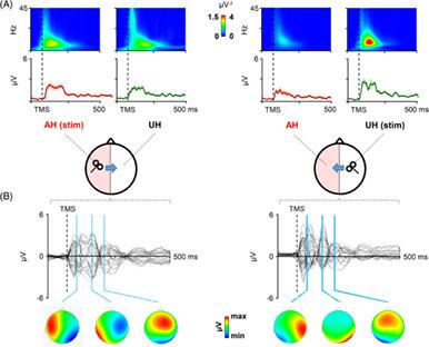
Interhemispheric interactions in stroke patients are frequently characterized by abnormalities, in terms of balance and inhibition. Previous results showed an impressive variability, mostly given to the instability of motor‐evoked potentials when evoked from the affected hemisphere. We aim to find reliable interhemispheric measures in stroke patients with a not‐evocable motor‐evoked potential from the affected hemisphere, by combining transcranial magnetic stimulation (TMS) and electroencephalography. Ninteen stroke patients (seven females; 61.26 ± 9.8 years) were studied for 6 months after a first‐ever stroke in the middle cerebral artery territory. Patients underwent four evaluations: clinical, cortical, corticospinal, and structural. To test the reliability of our measures, the evaluations were repeated after 3 weeks. To test the sensitivity, 14 age‐matched healthy controls were compared to stroke patients. In stroke patients, stimulation of the affected hemisphere did not result in any inhibition onto the unaffected. The stimulation of the unaffected hemisphere revealed a preservation of the inhibition mechanism onto the affected. This resulted in a remarkable interhemispheric imbalance, whereas this mechanism was steadily symmetric in healthy controls. This result was stable when cortical evaluation was repeated after 3 weeks. Importantly, patients with a better recovery of the affected hand strength were the ones with a more stable interhemispheric balance. Finally, we found an association between microstructural integrity of callosal fibers, suppression of interhemispheric TMS‐evoked activity and interhemispheric connectivity. We provide direct and sensitive cortical measures of interhemispheric imbalance in stroke patients. These measures offer a reliable means of distinguishing healthy and pathological interhemispheric dynamics.
中文翻译:

结合经颅磁刺激和脑电图揭示中风患者半球间不平衡的证据
中风患者的半球间相互作用通常以平衡和抑制方面的异常为特征。先前的结果显示出令人印象深刻的可变性,主要是由于从受影响的半球诱发时运动诱发电位的不稳定性。我们的目标是通过结合经颅磁刺激 (TMS) 和脑电图,在受累半球具有不可唤起的运动诱发电位的中风患者中找到可靠的半球间测量值。19 名中风患者(7 名女性;61.26 ± 9.8 岁)在大脑中动脉区域首次中风后接受了为期 6 个月的研究。患者接受了四项评估:临床评估、皮质评估、皮质脊髓评估和结构评估。为了测试我们的措施的可靠性,评估在 3 周后重复。为了测试灵敏度,将 14 名年龄匹配的健康对照者与中风患者进行了比较。在中风患者中,对受影响的半球的刺激不会导致对未受影响的半球的任何抑制。未受影响的半球的刺激揭示了对受影响的抑制机制的保留。这导致了显着的半球间不平衡,而这种机制在健康对照中稳定对称。当 3 周后重复皮质评估时,该结果是稳定的。重要的是,受影响的手部力量恢复得更好的患者是具有更稳定的半球间平衡的患者。最后,我们发现胼胝体纤维的微观结构完整性、抑制半球间 TMS 诱发的活动和半球间连接性之间存在关联。我们提供中风患者半球间失衡的直接和敏感的皮质测量。这些措施提供了区分健康和病理的半球间动态的可靠方法。
更新日期:2021-03-03
中文翻译:
结合经颅磁刺激和脑电图揭示中风患者半球间不平衡的证据
中风患者的半球间相互作用通常以平衡和抑制方面的异常为特征。先前的结果显示出令人印象深刻的可变性,主要是由于从受影响的半球诱发时运动诱发电位的不稳定性。我们的目标是通过结合经颅磁刺激 (TMS) 和脑电图,在受累半球具有不可唤起的运动诱发电位的中风患者中找到可靠的半球间测量值。19 名中风患者(7 名女性;61.26 ± 9.8 岁)在大脑中动脉区域首次中风后接受了为期 6 个月的研究。患者接受了四项评估:临床评估、皮质评估、皮质脊髓评估和结构评估。为了测试我们的措施的可靠性,评估在 3 周后重复。为了测试灵敏度,将 14 名年龄匹配的健康对照者与中风患者进行了比较。在中风患者中,对受影响的半球的刺激不会导致对未受影响的半球的任何抑制。未受影响的半球的刺激揭示了对受影响的抑制机制的保留。这导致了显着的半球间不平衡,而这种机制在健康对照中稳定对称。当 3 周后重复皮质评估时,该结果是稳定的。重要的是,受影响的手部力量恢复得更好的患者是具有更稳定的半球间平衡的患者。最后,我们发现胼胝体纤维的微观结构完整性、抑制半球间 TMS 诱发的活动和半球间连接性之间存在关联。我们提供中风患者半球间失衡的直接和敏感的皮质测量。这些措施提供了区分健康和病理的半球间动态的可靠方法。


 京公网安备 11010802027423号